Science Note
Mitochondrial Transfer: A New Mechanism in Cancer Cell Savival [July 1, 2025]
| Cancer cells can hijack mitochondria from immune cells to boost their metabolism and suppress immunity¹. Remarkably, a recent study also revealed that cancer cells transfer mutated mitochondria to lymphocytes, leading to dysfunction and senescence². Together, these findings illustrate how cancer cells exploit mitochondrial dynamics to create an immunosuppressive microenvironment. | ||||||||||||||||||||||||||
|
Immune evasion through mitochondrial transfer in the tumour microenvironment (Nature, 2025) Highlighted technique: Mitochondrial transfer and mitophagy are key experimental targets in this study, which evaluates these phenomena using mitochondrial detection probes, mitochondria-targeted fluorescent proteins, and lysosomal marker staining. Related technique Mitophagy Imaging without Transfection, Cellular Senescence Detection (Used in this article) |
||||||||||||||||||||||||||
|
Intercellular nanotubes mediate mitochondrial trafficking between cancer and immune cells (Nature Nanotechnology, 2021) Highlighted technique: In this study, nanotubes formed between cancer cells and immune cells were visualized using actin filament labeling, and the transfer of mitochondria through these structures was observed using mitochondrial probes. This mitochondrial transfer was further supported by the co-localized movement of labeled mitochondrial DNA (mtDNA) within the nanotubes. Related technique Mitochondrial Staining Dyes, OCR Assay, Glysolysis/OXPHOS Assay |
||||||||||||||||||||||||||
Related Techniques (click to open/close)
|
||||||||||||||||||||||||||
Application Note I (click to open/close)
|
||||||||||||||||||||||||||
|
|
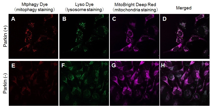
CCCP was added to Parkin-expressing HeLa cells and normal HeLa cells to confirm mitophagy. In the experiment, lysosomes and mitochondria were co-stained, and a strong mitophagic signal was observed on mitochondria and lysosomes in Parkin-expressing HeLa cells.
Products in Use |