MitoBright IM Red for Immunostaining
Mitochondria Fluorescent Probe for Immunostaining Red
- Capable of co-stained with immunostaining
- Higher retention in mitochondria after fixation & membrane permeabilization
-
Product codeMT15 MitoBright IM Red for Immunostaining
| Unit size | Price | Item Code |
|---|---|---|
| 20 μl x 1 | $157.00 | MT15-10 |
| 20 μl x 3 | $350.00 | MT15-12 |

Description
Cells have intracellular organelles that function to produce energy, synthesize and degrade proteins. Typical organelles include the nucleus, mitochondria, endoplasmic reticulum, and lysosomes. Although research on the functions of these organelles has been actively conducted for some time, recent developments in imaging technology, such as super-resolution imaging, have revealed that not only the functions of each organelle but also the coordination among organelles is important. One such example is the mitochondria-associated endoplasmic reticulum membrane, which is the interface between mitochondria and the endoplasmic reticulum (ER). Visualization of mitochondria is necessary to observe the interaction between mitochondria and other organelles. Fluorescent protein labeling, immunostaining, and mitochondria-selective small molecule fluorochromes are commonly used for this purpose, although these methods have problems. The fluorescent protein labeling method requires gene transfer, and multiple staining may not be able to perform in the immunostain method depending on the animal species of the primary antibody. In addition, commercially available mitochondria-selective small molecule fluorescent dyes do not have good retention properties and are difficult to use in immunostaining.
To overcome the problems above, Dojindo has developed a new mitochondria-selective small-molecule fluorescent dye, MitoBright IM Red, which covalently binds to proteins and other biomolecules. MitoBright IM Red, therefore, is able to simultaneously perform the staining mitochondria and the immunostaining.


Manual
Technical info
The existing molecular probe for mitochondrial staining has a problem with clarity when co-stained with immunostaining methods. MitoBright IM has a structure that allows it to be easily retained in mitochondria even during the immunostaining process after staining live cells. MitoBright IM is a new reagent that overcomes the problem of combined use with immunostaining because it has a structure that is easily retained in mitochondria.
Using MitoBright IM stained HeLa cells' mitochondria and then after fixation and membrane permeanbility treatment, co-stained with immunostaining using KDEL antibody, a marker protein of the endoplasmic reticulum(ER). The fluorescence intensity was measured in the area indicated by the blue arrow in the fluorescence image (right). As a result, the morphology of the mitochondria and the neighboring endoplasmic reticulum was clearly observed.
.png)
<Imaging Conditions>
MitoBright IM Red (Red) Ex: 561 nm, Em: 560-620 nm
KDEL antibody-Alexa 488 (Green) Ex: 488 nm, Em: 490-550 nm
Scale bar:10 μm
Feature of MitoBright IM Red
Common molecular probe for mitochondrial staining has a problem with retention to mitochondria when co-stained with immunostaining methods. MitoBright IM has a structure with improved retention properties compared to existing small molecule dyes, which reduces the loss of fluorescence intensity and localization during immunostaining.
・Comparison of Fluorescent Intensity
Using MitoBright IM and company T's existing reagent stain live cells followed by fixation and membrane permeability treatment. The below image shows the fluorescent intensity difference after each step. Although the fluorescence intensity of both reagents decreased after fixation and membrane permeability treatment compared to live cells. However, it was confirmed that MitoBright IM has higher retention and clarity than the existing reagent.
.png)
<Imaging Conditions>
Ex=561 nm, Em=560-620 nm
・Comparison of Clarity
The mitochondrial localization of MitoBright IM or existing reagents after immunostaining was compared using TOM20 antibody, a mitochondrial marker. HeLa cells were stained with MitoBright IM or existing reagents and then the fluorescence was observed by secondary antibody staining using TOM20 antibody. As a result, it was confirmed that the existing reagent dispersed outside of the mitochondria, resulting in a high background, whereas MitoBright IM suppressed the background and its localization was consistent with that of the TOM20 antibody.
.png)
.png)
<Imaging Conditions>
Ex=561 nm, Em=560-620 nm
Scale bar: 10 µm
Comparison with Existing Reagent
Unlike the MitoBright LT, the MitoBright IM is specialized for immunostaining applications. As same as MitoBright LT, the MitoBright IM is dissolved in DMSO and can be used immediately without any preparation step of staining solution. For other performance differences, please refer to the table below.
.png)
General Number of Usable Assays
| MitoBright IM Unit size | Fluorescent Microscopy | |
|---|---|---|
| (35 mm dish, 2 ml working solution/dish) | ||
| 20 μl | 10 dish | |
| 20 µl x3 | 30 dish | |
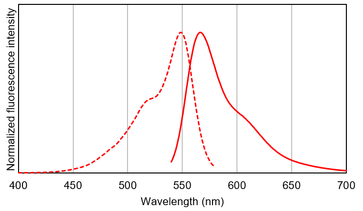
Fluorescent Spectrum
MitoBright IM Red
|
Q & A
-
Q
Is MitoBright IM Red DMSO solution stable after repeated freezing and thawing?
-
A
We have confirmed that 5 times of frozen and thawing the MitoBright IM Red DMSO solution is still stable. However, we recommend dividing the solution for long-term storage.
-
Q
When cells were stained with MitoBright IM after drug treatment, the staining was uneven. What can I do to improve it?
-
A
Please stain the cells with MitoBright IM before drug treatment.
Mitobright IM accumulates in mitochondria via mitochondrial membrane potential, if the drug treatment significantly decreases membrane potential, Mitobright IM can not accumulate in mitochondria.(For your references)
HeLa cells were stained with MitoBright IM before and after FCCP treatment and observed.
Fluorescence was observed when the cells were stained before FCCP treatment, but no fluorescence was observed when the cells were stained after FCCP treatment.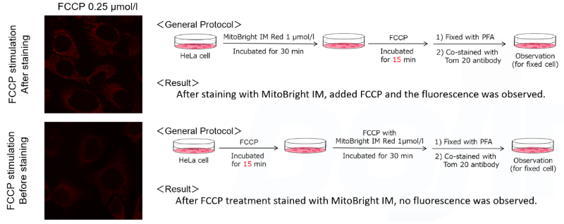
Handling and storage condition
| Keep tightly closed. Store in a deep freeze at -20℃, dark and dry place. |
Related products
The following related products are also used in research related to this product.