Mito-FerroGreen
Mitochondrial Iron Detection
-
Product codeM489 Mito-FerroGreen
-
CAS No.-
| Unit size | Price | Item Code |
|---|---|---|
| 1 set (50 ug x 2) | $322.00 | M489-10 |
*1 set (50 µg x 2) can be used for 10 assays at 35 mm dish (final concentration 5 µmol/L)

Description
Product Description
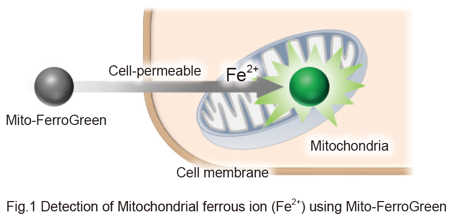
It is reported that iron is the most abundant transition metal element within an organism and shows various physiological activities. Recently, free iron in living cells is getting attention because its high reactivity is suggested to be related to cellular damage or death. Free iron exists in its stable redox states, ferrous ion (Fe2+) and ferric ion (Fe3+). In living cells, it is considered that understanding the behavior of Fe2+ is more important than that of Fe3+ because of the intracellular reductive environment, metal transporters and water solubility of Fe2+. Mito-FerroGreen is a novel fluorescent probe for the detection of ferrous ion (Fe2+) in mitochondria where Fe-S clusters and heme proteins are synthesized, and enables live cell fluorescent imaging of intracellular Fe2+. Mito-FerroGreen has no no chelating ability. Mito-FerroGreen and Fe2+ react irreversibly, which is different from the detection principle of calcium-ion probes such as Fluo 3.
| Product Name | Target | Detection Properties |
|---|---|---|
| FerroOrange | Intracellular Fe2+ | Microscopy, Plate reader Ex: 543 nm / Em: 580 nm |
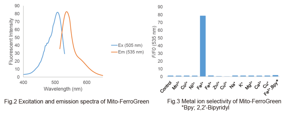
| Mito-FerroGreen | Mitochondrial Fe2+ | Microscopy Ex: 505 nm / Em: 535 nm |
| Lyso-FerroRed | Lysosomal Fe2+ | Microscopy, FCM, Plate reader Ex: 551 nm / Em: 571 nm |
| Iron Assay Kit -Colorimetric- | Fe2+ and Fe3+ | Plate reader Colorimetric, λ: 593 nm |
| Liperfluo | Lipid Peroxide | Microscopy, FCM Ex: 488 nm / Em: 500-550 nm |
| Lipid Peroxidation Probe -BDP 581/591 C11- |
Lipid Peroxidation Process |
Microscopy, FCM, Plate reader |
| MDA Assay Kit | Malondialdehyde | Plate reader Fluorescence, Ex: 540 nm / Em: 590 nm Colorimetric, λ: 532 nm |
| Cystine Uptake Assay Kit | Cystine uptake | Plate reader Ex: 485 nm / Em: 535 nm |
| GSSG/GSH Quantification Kit II | GSSG and GSH | Plate reader Colorimetric, λ: 405 nm |


Manual
Technical info
Detection of Mitochondrial ferrous iron (Fe2+) using Mito-FerroGreen
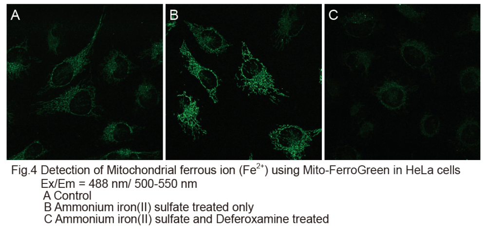
1. HeLa cells were seeded on μ-slide 8 well (ibidi) and cultured at 37oC overnight in a 5% CO2 incubator.
2. The cells were washed with HBSS three times.
3. Mito-FerroGreen working solution (5 μmol/l, 200 μl) were added to the cells, and the cells were incubated at 37oC for 30 minutes in a 5% CO2 incubator.
4. After supernatant was discarded, 10 mmol/l deferoxamine mesylate salt (sigma) prepared with HBSS was added to the cells, and the cells were incubated at 37oC for 30 minutes in a 5% CO2 incubator.
5. The supernatant was discarded and the cells were washed with HBSS three times. After the HBSS was removed, serum-free medium was added to the cells.
6. Ammonium iron(II) sulfate prepared with purified water (10 mmol/l) was added to the cell to be the final concentration of 100 μmol/l.
7. The cells were incubated at 37oC for 1 hour in a 5% CO2 incubator, and the cells were washed with HBSS three times.
8. The cells were observed by confocal fluorescence microscopy.
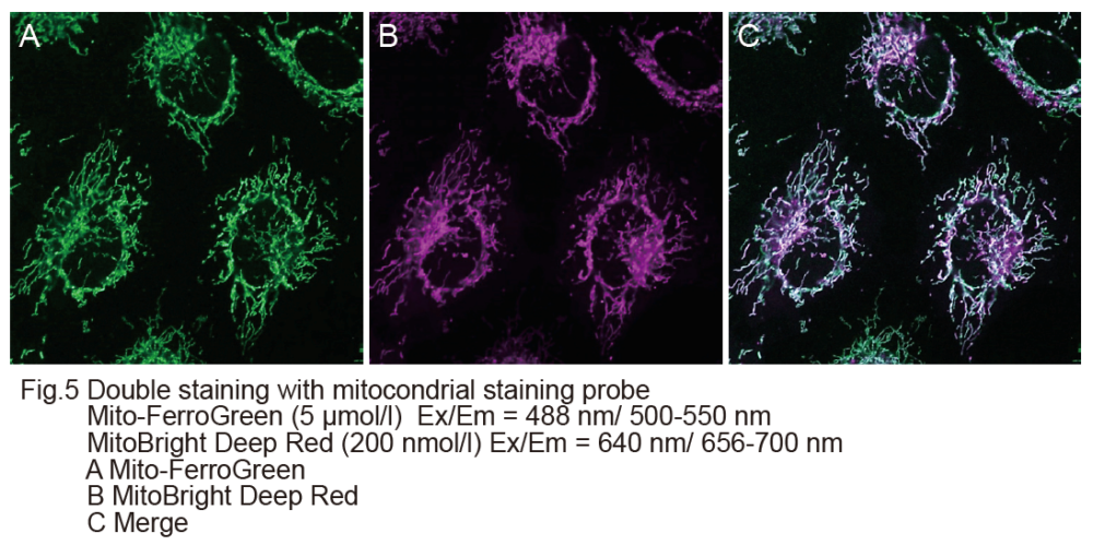
Double staining with mitochondrial staining probe
1. HeLa cells were seeded on μ-slide 8 well (ibidi) and cultured at 37oC overnight in a 5% CO2 incubator.
2. The cells were washed with HBSS three times and the supernatant was removed.
3. HBSS (200 μl) containing the final concentration of 5 μmol/l Mito-FerroGreen and 200 nmol/l MitoBright Deep Red (Dojindo※) were added to the cells and the cells were incubated at 37oC for 30 minutes in a 5% CO2 incubator.
4. The cells were washed with HBSS three times.
5. After the HBSS was removed, 200 μl of serum-free medium was added to the cells, and 10 mmol/l ammonium iron(II) sulfate prepared with purified water was added to be the final concentration of 100 μmol/l.
6. The cells were incubated at 37oC for 1 hour in a 5% CO2 incubator, and the cells were washed with HBSS three times.
7. The cells were observed by confocal fluorescence microscopy.
※Our sincere apology that we discontinued distributing MitoBright Deep Red.
Please refer to MitoBright LT Deep Red (Code#:MT12) which improved retention ability.
References
1) T. Hirayama, S. Kadota, M. Niwa and H. Nagasawa, "A mitochondria-targeted fluorescent probe for selective detection of mitochondrial labile Fe(II)", Metallomics., 2018, DOI: 10.1039/C8MT00049B, ※「Mito-FerroGreen」 =「Ac-MtFluNox」.
2) T. Issitt, E. Bosseboeuf, N. Winter, N. Dufton, G. Gestri, V. Senatore, A. Chikh, A. Randi, C. Raimondi, "Neuropilin-1 controls endothelial homeostasis by regulating mitochondrial function and iron-dependent oxidative stress via ABCB8", iScience., 2018,DOI: 10.1016/j.isci.2018.12.005 .
3) E. E. Mon, F. Y. Wei, R. N. R. Ahmad, T. Yamamoto, T. Moroishi and K. Tomizawa, "Regulation of mitochondrial iron homeostasis by siderofexin 2 ", J Physiol Sci., 2018,doi:10.1007/s12576-018-0652-2.
4) M. Fujimaki, N. Furuya, S. Saiki, T. Amo, Y. Imamichi and N. Hattori, "Iron supply via NCOA4-mediated ferritin degradation maintains mitochondrial functions", Mol. Cell. Biol.., 2019,doi: 10.1128/MCB.00010-19.
5) K. Tomita, M. Fukumoto, K. Itoh, Y. Kuwahara, K. Igarashi, T. Nagasawa, M. Suzuki, A. Kurimasa and T. Sato, "MiR-7-5p is a key factor that controls radioresistance via intracellular Fe2+ content in clinically relevant radioresistant cells.", Biochem Biophys Res Commun.., 2019,doi: 10.1016/j.bbrc.2019.08.117.
6) Y. Wang and M. Tang, "PM2.5 induces ferroptosis in human endothelial cells through iron overload and redox imbalance", Environ. Pollut., 2019, 264, doi: 10.1016/j.envpol.2019.07.105.
7) KF. Yambire, C. Rostosky, T. Watanabe, D. Pacheu-Grau, S. Torres-Odio,A. Sanchez-Guerrero,O. Senderovich, EG. Meyron-Holtz,I.Milosevic, J. Frahm, AP. West and N. Raimundo, "Impaired lysosomal acidification triggers iron deficiency and inflammation in vivo.", Elife, 2019, 3, (8), doi:10.7554/eLife.51031.
8) H. Nishizawa, M. Matsumoto, T. Shindo, D. Saigusa, H. Kato, K. Suzuki, M. Sato, Y. Ishii, H. Shimokawa and K. Igarashi, "Ferroptosis is controlled by the coordinated transcriptional regulation of glutathione and labile iron metabolism by the transcription factor BACH1", J. Biol. Chem., 2019,doi: 10.1074/jbc.RA119.009548.
9)Y. akashima, A. Hayano and B. Yamanaka, Metabolome analysis reveals excessive glycolysis via PI3K/AKT/mTOR and RAS/MAPK signaling in methotrexate-resistant primary CNS lymphoma-derived cells.", Clin. Cancer Res., 2020, DOI:10.1158/1078-0432.
Handling and storage condition
| Purity (HPLC): | ≧ 90.0 % |
|---|---|
| Fluorescence spectrum: | To pass test |
| -20°C, Protect from light, Nitrogen substitution, Protect from moisture |
Related products
The following related products are also used in research related to this product.