 Hidden sections will not be printed.
General Information
It is reported that iron is the most abundant transition metal element within an organism and shows various physiological activities. Recently, free iron in living cells is getting attention because its high reactivity is suggested to be related to cellular damage or death. Free iron exists in its stable redox states, ferrous ion (Fe2+) and ferric ion (Fe3+).
In living cells, it is considered that understanding the behavior of Fe2+ is more important than that of Fe3+ because of the intracellular reductive environment, metal transporters and water solubility of Fe2+. Mito-FerroGreen is a novel fluorescent probe for the detection of ferrous ion (Fe2+) in mitochondria where Fe-S clusters and heme proteins are synthesized, and enables live cell fluorescent imaging of intracellular Fe2+.
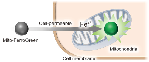
Figure 1 Detection of mitochondrial ferrous ion (Fe2+) using Mito-FerroGreen
-
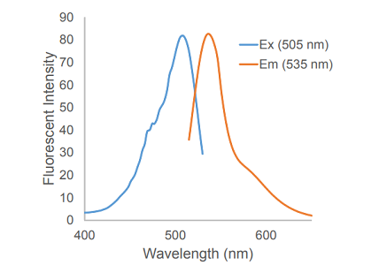
Figure 2 Excitation and emission spectra of Mito-FerroGreen
-
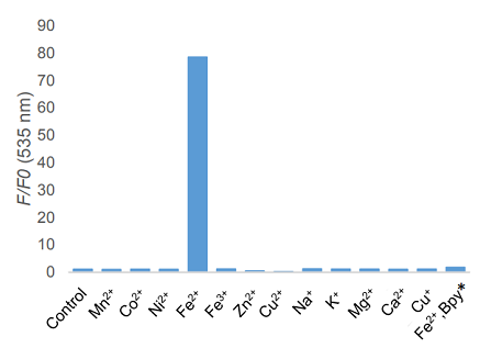
Figure 3 Metal ion selectivity of Mito-FerroGreen
*Bpy; 2,2'-Bipyridyl
Contents
| Mito-FerroGreen | 50 µg x 2 |
Storage Condition
- Store at -20oC and protect from light.
Required Equipment and Materials
- Dimethyl sulfoxide (DMSO) or Ethanol
- HBSS
- Serum-free medium
- Micropipettes
Preparation of Solution
Preparation of Mito-FerroGreen working solution
Add 53 μl of DMSO to a tube containing of 50 μg of Mito-FerroGreen and dissolve it by pipetting to prepare 1 mmol/l Mito-FerroGreen solution. Dilute the 1mmol/l Mito-FerroGreen solution with HBSS to prepare 5 μmol/l Mito-FerroGreen working solution.
- 1 mmol/l Mito-FerroGreen DMSO solution and 5 μmol/l Mito-FerroGreen working solution are unstable. Please prepare the solutions just before the staining experiment and use them immediately. Do not use the rest of the Mito-FerroGreen solutions due to the increase of background signal resulted from degradation of the Mito-FerroGreen solution. Ethanol can also be used for preparation. The Ethanol solution is stable at -20oC for 2 weeks.
- Substitute serum-free medium for HBSS if needed. However, please note that serum-containing medium cannot be used due to causing high background.
General Protocol
- Inoculate cells to a dish for assay and culture the dish at 37oC in a 5% CO2 incubator.
- Discard the supernatant and wash the cells with HBSS or serum-free medium three times.
- Add 5 μmol/l Mito-FerroGreen working solution to the cells and incubate at 37oC for 30 minutes in a 5% CO2 incubator.
- Discard the supernatant and wash the cells with HBSS or serum-free medium three times.
- Add medium containing stimulating agents and incubate at 37oC in a 5% CO2 incubator.
- Please optimize the incubation time according to stimulating conditions.
- Observe cells by fluorescence microscopy.
- The order of step 3 and step 5 can be changed in response to experimental conditions, i.e. 5 μmol/l Mito-FerroGreen working solution can be added to the cells after agent stimulation.
Experimental Example 1
Detection of mitochondrial ferrous iron (Fe2+) using Mito-FerroGreen.
- HeLa cells (2.0×10⁴ cells/well) were seeded on μ-slide 8 well (ibidi) and cultured at 37 oC overnight in a 5% CO2 incubator.
- The cells were washed with HBSS (200 μl) three times.
- Mito-FerroGreen working solution (5 μmol/l, 200 μl) were added to the cells, and the cells were incubated at 37oC for 30 minutes in a 5% CO2 incubator.
- After supernatant was discarded, 10 mmol/l deferoxamine mesylate salt (sigma) prepared with HBSS (200 μl) was added to the cells, and the cells were incubated at 37oC for 30 minutes in a 5% CO2 incubator.
- The supernatant was discarded and the cells were washed with HBSS (200 μl) three times. After the HBSS was removed, serum-free medium (200 μl) was added to the cells.
- Ammonium iron (II) sulfate (10 mmol/l) was prepared with purified water.
- Ammonium iron (II) sulfate (2 μl) was added to wells (The final concentration:100 μmol/l). To mix Ammonium iron (II) sulfate and serum-free medium, the entire medium was pipetted up from wells and then immediately pipetted back one time.
- *Please do not disturb the cells during pipetting.
- *When adding 10 mmol/l Ammonium iron (II) sulfate to well, please exactly follow step 7 as described. Do not add pre-prepared 100 μmol/l Ammonium iron (II) sulfate to cells. It may result in precipitation of Ammonium iron (II) sulfate during the experiment due to a vortex or a pipetting.
- The cells were incubated at 37oC for 1 hour in a 5% CO2 incubator, and the cells were washed with HBSS (200 μl) three times.
- The cells were observed by confocal fluorescence microscopy.
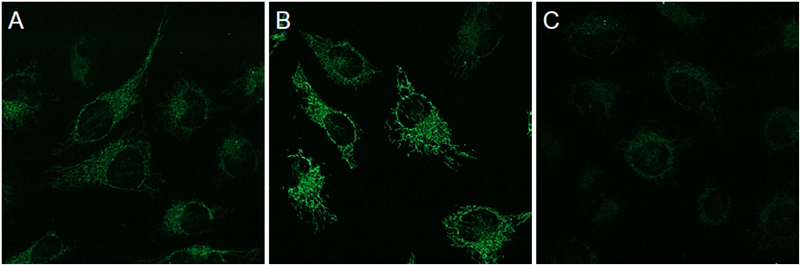
Figure 4 Detection of mitochondrial ferrous ion (Fe2+) using Mito-FerroGreen in HeLa cells
Ex/Em = 488 nm/ 500-550 nm A Control
B Ammonium iron (II) sulfate treated only
C Ammonium iron (II) sulfate and Deferoxamine treated
Experimental Example 2
Double staining with mitochondrial staining probe
- HeLa cells (2.0×10⁴ cells/well) were seeded on μ-slide 8 well (ibidi) and cultured at 37 oC overnight in a 5% CO2 incubator.
- The cells were washed with HBSS (200 μl) three times.
- HBSS (200 μl) containing the final concentration of 5 μmol/l Mito-FerroGreen and 200 nmol/l MitoBright Deep Red (Dojindo, Code: MT08) were added to the cells and the cells were incubated at 37oC for 30 minutes in a 5% CO2 incubator.
- The supernatant was discarded and the cells were washed with HBSS (200 μl) three times. After the HBSS was removed, serum-free medium (200 μl) was added to the cells.
- Ammonium iron (II) sulfate (10 mmol/l) was prepared with purified water.
- Ammonium iron (II) sulfate (2 μl) was added to wells (The final concentration:100 μmol/l). To mix Ammonium iron (II) sulfate and serum-free medium, the entire medium was pipetted up from wells and then immediately pipetted back one time.
- Please do not disturb the cells during pipetting.
- When adding 10 mmol/l Ammonium iron (II) sulfate to well, please exactly follow step 6 as described. Do not add pre-prepared 100 μmol/l Ammonium iron (II) sulfate to cells. It may result in precipitation of Ammonium iron (II) sulfate during the experiment due to a vortex or a pipetting.
- The cells were incubated at 37oC for 1 hour in a 5% CO2 incubator, and the cells were washed with HBSS (200 μl) three times.
- The cells were observed by confocal fluorescence microscopy.
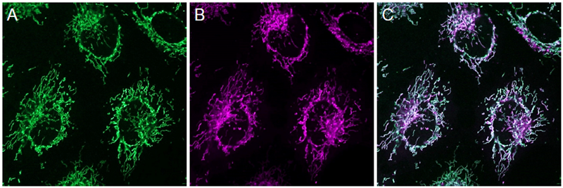
Figure 5 Double staining with mitochondrial staining probe
Mito-FerroGreen (5 μmol/l) Ex/Em = 488 nm/ 500-550 nm
MitoBright Deep Red (200 nmol/l) Ex/Em = 640 nm/ 656-700 nm
A Mito-FerroGreen
B MitoBright Deep Red
C Merge
Frequently Asked Questions / Reference


M489: Mito-FerroGreen
Revised Aug., 22, 2023