


LysoPrime Deep Red - High Specificity and pH Resistance
Lysosome Staining Deep Red
- High specificity for lysosomes
- pH resistance
- Able to stain lysosome overnight
-
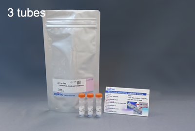
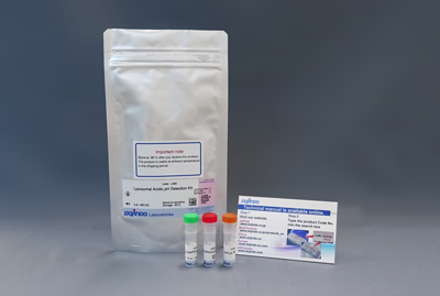
Product codeL264 LysoPrime Deep Red - High Specificity and pH Resistance
| Unit size | Price | Item Code |
|---|---|---|
| 1 tube | $243.00 | L264-10 |
| 3 tube | $511.00 | L264-12 |
35 mm dish ×10, μ-Slide 8 well ×10, 96-well Plate ×2 for each tube.


Description
The lysosome is an organelle in which a biomembrane forms an acid vacuole. Lysosomes contain various degrading enzymes and contribute to maintaining intracellular homeostasis by acting as a waste disposal system. Recent findings reveal that lysosomal dysfunction is related to some neurodegenerative disorders. Consequently, the investigation of lysosomal function is attracting considerable interest in the scientific community.
Many small fluorescent probes are used to monitor lysosomes in living cells. Dojindo’s LysoPrime Deep Red overcomes known problems with fluorescent lysosome probes, such as lack of specificity for lysosomes and staining dependent on the lysosomal pH. In addition, the high-retentivity of LysoPrime Deep Red enables long-term imaging experiments.
Lysosomal Analysis Products
| Product Name | Lysosomal pH Detection Dyes and Fluorescence Properties |
Lysosomal Quantity Detection Dyes and Fluorescence Properties |
|---|---|---|
| Lysosomal Acidic pH Detection Kit | pHLys Red Ex: 561 nm / Em: 560-650 nm |
LysoPrime Green Ex: 488 nm / Em: 500-600 nm |
| Lysosomal Acidic pH Detection Kit - Green/Deep Red | pHLys Green Ex: 488 nm / Em: 490-550 nm |
LysoPrime Deep Red Ex: 633 nm / Em: 640-700 nm |
| pHLys Red - Lysosomal Acidic pH Detection | pHLys Red Ex: 561 nm / Em: 560-650 nm |
|
| LysoPrime Deep Red - High Specificity and pH Resistance | LysoPrime Deep Red Ex: 633 nm / Em: 640-700 nm |
|
| LysoPrime Green- High Specificity and pH Resistance | LysoPrime Green Ex: 488 nm / Em: 500-600 nm |


Manual
Technical info
Comparative Performance Analysis
All data were generated in-house under identical imaging and treatment conditions.
Lysosome tracking dye
| Dye | LysoPrime Green | LysoPrime Deep Red |
Lysosomal Tracker Green |
|
pH Stability |
Resistant to pH elevation |
Resistant to pH elevation |
Reduced under pH elevation |
|
Lysosomal Retention Time |
Long-term Retention |
Long-term Retention |
Rapid Signal Decline |
High Specificity for Lysosomes
The lysosomal localization of competitor's dye and LysoPrime Deep Red was compared using HeLa cells expressing the lysosomal marker protein LAMP1-GFP. The LysoPrime Deep Red localized better to lysosomes than the existing dye (Merged image), proved that LysoPrime Deep Red has better localization to lysosomes than existing dyes.

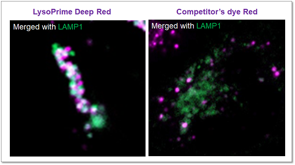
Green: Ex= 488 nm, Em= 500-570 nm
Deep Red : Ex= 633 nm, Em= 640-700 nm
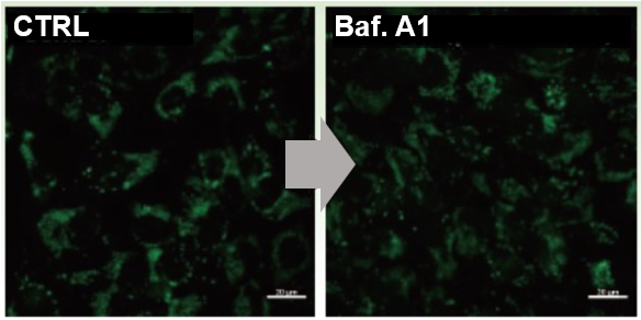
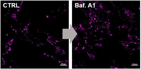
pH Resistance
LysoPrime Deep Red and other competitor's dyes can accumulate in acidic lysosomes. However, after being treated with Bafilomycin A1 (Baf. A1), a lysosomal acidity inhibitor, the competitor's dyes' fluorescent signal was significantly reduced when the lysosome is changed from acidic to neutral. because the dyes were leaked from lysosomes. In the meanwhile, LysoPrime Deep Red was retained in lysosomes, resulting in a less decrease in fluorescence signal and clearer results compared to the competitor's dye.
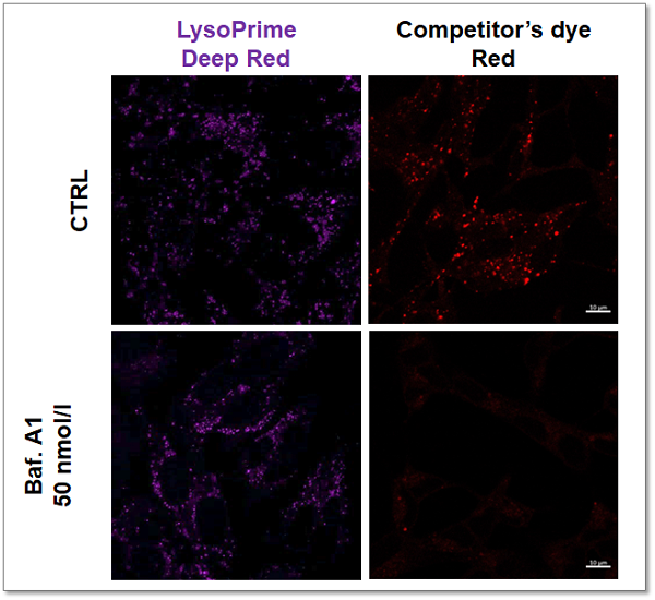

<Experimental Conditions>
Deep Red: Ex= 633 nm, Em= 640-700 nm
Red: Ex= 561 nm, Em= 560-620 nm
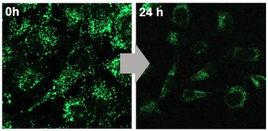
High Retention in Lysosome
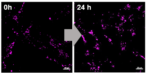
The lysosomal retention ability is compared using LysoPrime Deep Red stained cells and competitor's dye stained cells. The result showed that, after 24 hours of staining, the fluorescence intensity of the competitor's dye was decreased obviously. On the other hand,
LysoPrime Deep Red maintained its fluorescence intensity.

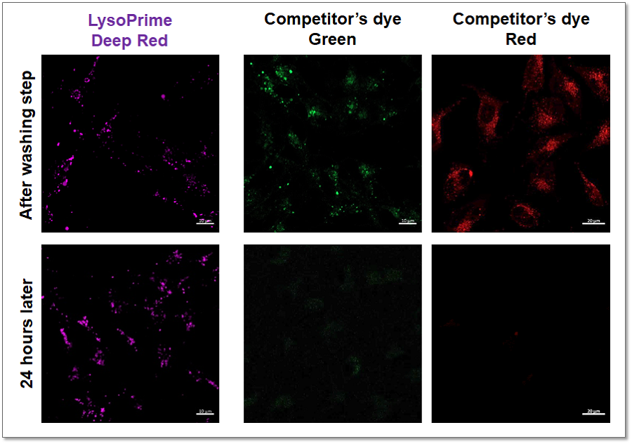
<Experimantal Conditions>
Deep Red: Ex= 633 nm, Em= 640-700 nm
Green: Ex= 488 nm, Em= 490-550 nm
Red: Ex= 561 nm, Em= 560-620 nm
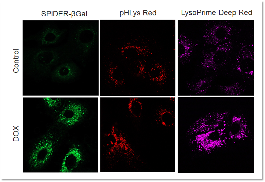
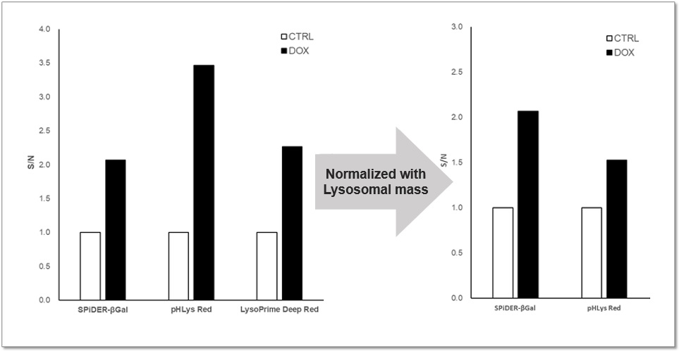
Application Data: Lysosomal Mass and pH Exchange in Senescence-induced Cells
We analyzed senescence-associated acidic β-galactosidase (SA-βGal) activity and lysosomal mass and pH in A549 cells treated with Doxorubicin (DOX) to induce senescence. The SA-βGal activity was detected by SG03 Cellular Senescence Detection Kit - SPiDER-βGal, the lysosomal mass and pH were detected separately with LysoPrime Deep Red and L265 pHLys Red. Fluorescence imaging showed that the increase in lysosomal mass and pH acidification were observed in senescence-induced cells, and the normalized fluorescence intensity of lysosomal mass and pH by plate reader measurement showed the same result.

<Experimental Conditions>
SA-βGal(Green):
Ex = 488 nm, Em = 490 – 550 nm
Lysosomal pH (Red):
Ex = 561 nm, Em = 560 – 620 nm
Lysosomal mass (Deep Red):
Ex = 633 nm, Em = 640 – 700 nm
<Experimental Conditions>
SA-βGal: Ex = 525 – 535 nm, Em = 550 – 570 nm
Lysosomal pH: Ex = 555 – 565 nm, Em = 590 – 610 nm
Lysosomal mass: Ex = 645 – 655 nm, Em = 690 – 710 nm
References
| No. | Cell Type | Inducer | Instrument | Reference (Link) |
|---|---|---|---|---|
| 1) | Human astrocytes | HIV-1 Tat, Mutant Tat | Microscope | N. Rezagholizadeh, G. Datta, W.A. Hasler, E.C. Nguon, E.V. Smokey, N. Khan, X. Chen, "SLC38A9 is directly involved in Tat-induced endolysosome dysfunction and senescence in astrocytes", Life Sci Alliance, 2025, doi:10.26508/lsa.202503231 |
| 2) | Human astrocytes | TLR7 agonist R837, Tat protein | Microscope | N. Rezagholizadeh, G. Datta, W.A. Hasler, E.C. Nguon, E.V. Smokey, X. Chen, "TLR7 Mediates HIV-1 Tat-Induced Cellular Senescence in Human Astrocytes", Aging Cell, 2025, doi:10.1111/acel.70086 |
Q & A
-
Q
Can I use medium with serum to staining the cells?
-
A
LysoPrime Deep Red is affected by serum, please prepare the working solution with serum-free medium or HBSS.
-
Q
When should I add the stimulation? Before the LysoPrime Deep Red staining or after?
-
A
Please add the stimulation after the LysoPrime Deep Red staining. Although LysoPrime Deep Red can remain in lysosomes even if the pH changes in lysosomes. If you add the stimulation before the staining, LysoPrime Deep Red cannot remain in lysosomes if the pH has been changed to near neutral due to drug stimulation.
-
Q
I want to evaluate the lysosomal mass and pH at the same time, can I use pHLys Red to co-staining?
-
A
Yes, you can. However, please notice that you should stain the cells with LysoPrime Deep Red first, and then stain with pHLys Red.
-
Q
Which instruments is applicable for LysoPrime Deep Red?
-
A
LysoPrime Deep Red is applicable for Fluorescent microscopes, Confocal microscopes, and plate readers.
・Confocal microscope
Ex = 633 nm, Em = 640-700 nm・Fluorescent microscope
Cy5 Filter
Ex = 590 - 650 nm, Em = 660 - 740 nm・Plate reader
Ex = 645 – 655 nm, Em = 690 – 710 nm
-
Q
What is the recommended final concentration of LysoPrime Deep Red?
-
A
We recommend using LysoPrime Deep Red at a 1,000x dilution, but if you want to optimize the staining conditions, please refer to the following range.
Use a dilution between 2,000x and 5,000x.
-
Q
The fluorescence of LysoPrime Deep Red decreased after drug treatment. How can I improve this?
-
A
We recommend considering a reduction in drug concentration or exposure time. If they do not resolve the issue, we recommend using LysoPrime Green as an alternative.
At a concentration 20 times higher than the recommended concentration of Bafilomycin A1 (1000nM vs 50nM), LysoPrime Green demonstrated higher retention within lysosomes than LysoPrime Deep Red, as it binds directly to lysosomal proteins.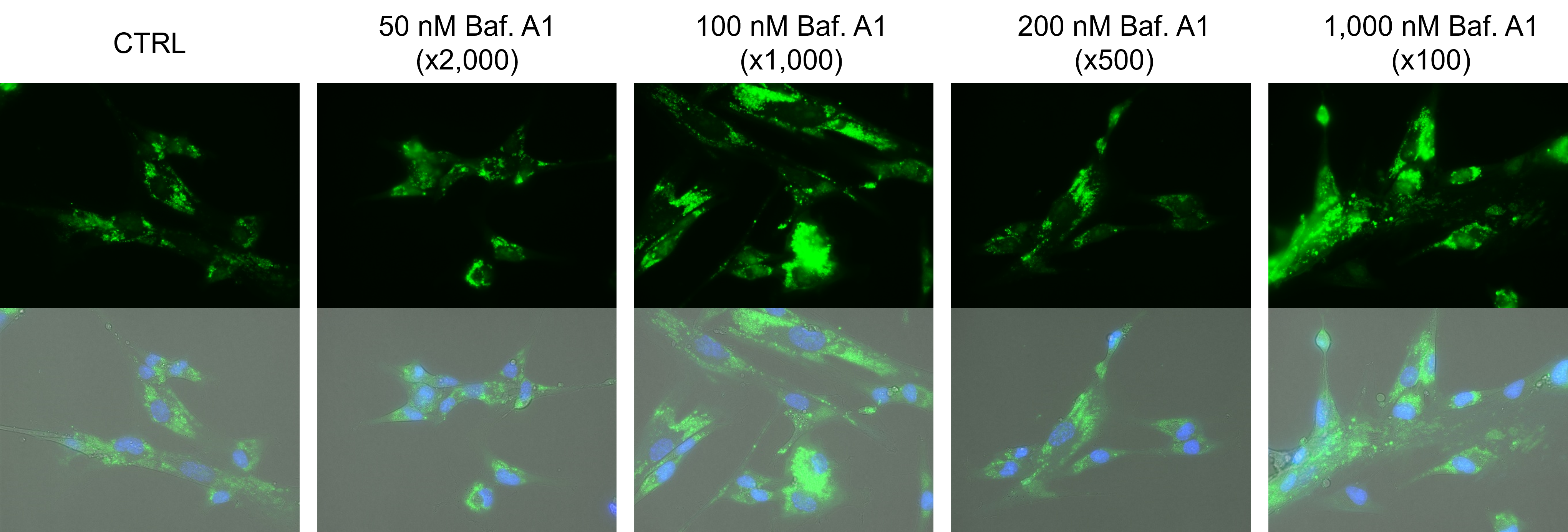
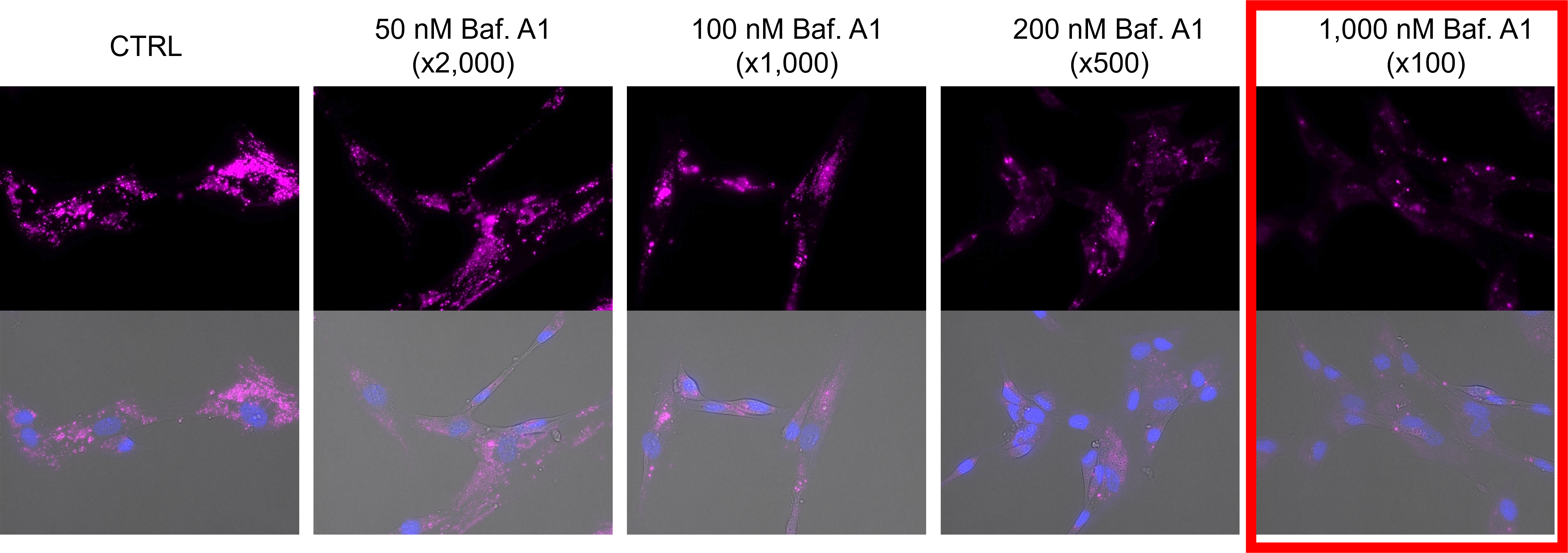
Comparison of LysoPrime Green / Deep Red retention at different bafilomycin A1 concentrations
• LysoPrime Green: recommended concentration x2,000 (recommended concentration range: x1,000 – 4,000)
• LysoPrime Deep Red: recommended concentration x1,000 (recommended concentration range x1,000–5,000)
• Bafilomycin A1: recommended concentration x2,000 (recommended concentration range x1,000–5,000)
-
Q
Can cells be fixed after staining with LysoPrime Deep Red?
-
A
Fixation is not recommended.
Handling and storage condition
| Appearance: | Purple Solid |
|---|---|
| Dye content: | To pass test |
| 1. Storage: -20℃; 2. Moisture-proof |
Related products
The following related products are also used in research related to this product.
-
ROS Detection
ROS Assay Kit -Photo-oxidation Resistant DCFH-DA-
-
Endocytosis Detection
ECGreen-Endocytosis Detection
-
Lysosome Staining Dye Green
LysoPrime Green - High Specificity and pH Resistance
-
Cell Membrane Staining
PlasMem Bright Green
-
Lysosomal pH Detection Reagent Red
pHLys Red - Lysosomal Acidic pH Detection
-
Lysosomal Acidic pH Detection Kit - Green/Red
Lysosomal Acidic pH Detection Kit


