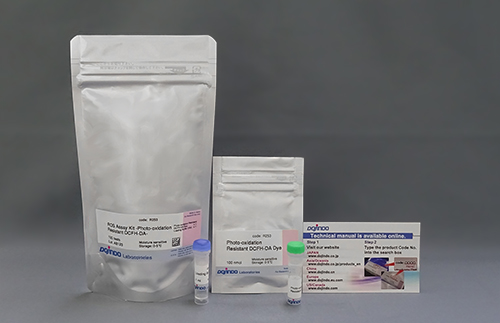
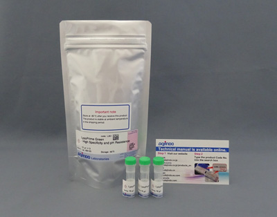
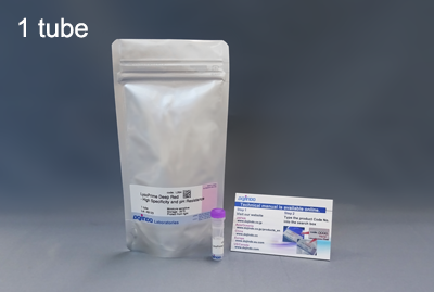
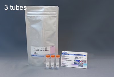
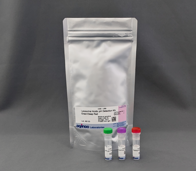
Lysosomal Acidic pH Detection Kit
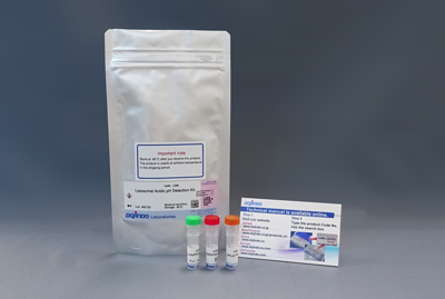
Lysosomal Acidic pH Detection Kit - Green/Red
- High Sensitivity
- High Specificity
- All-in-one Kit with Lysosome Acidification Inhibitor included
-
Product codeL266 Lysosomal Acidic pH Detection Kit
| Unit size | Price | Item Code |
|---|---|---|
| 1 set | $388.00 | L266-10 |
35 mm dish ×10, μ-Slide 8 well ×10, 96-well Plate ×2 for each tube.
<Storage Precautions>
・For long-term storage, storage below -80°C or in liquid nitrogen is recommended.
・Even store under -20°C conditions may cause degradation over time.
If storing below -80°C is difficult, please consider using the reagent as soon as possible.
・Please avoid repeated freezing and thawing, as it may cause degradation.
*Also see [L268] Lysosomal Acidic pH Detection Kit - Green/Deep Red, this product that can be stored at -20°C.
| 1 set | LysoPrime Green pHLys Red Bafilomycin A1 |
x 1 x 1 x 1 |
|---|
Description
The lysosome is an organelle in which a biomembrane forms an acid vacuole. Lysosomes contain various degrading enzymes and contribute to maintaining intracellular homeostasis by acting as a waste disposal system. Recent findings reveal that lysosomal dysfunction is related to some neurodegenerative disorders. Consequently, the investigation of lysosomal function is attracting considerable interest in the scientific community.
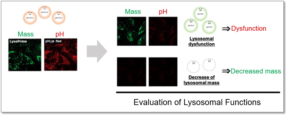
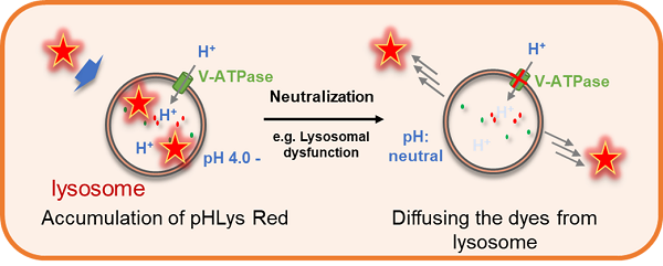
This kit includes lysosome staining dyes, pHLys Red (pH-sensitive), and LysoPrime Green (pH-resistant). The pHLys Red and LysoPrime Green accumulate in the intact lysosomes. The fluorescence intensity of pHLys Red is enhanced as the acidity increases, and weak fluorescence is observed when lysosomes are neutralized due to the lysosomal dysfunction. On the other hand, LysoPrime Green gives stable emissions even lysosomes are neutralized. Lysosomal pH and lysosomal mass can be measured by combining these two dyes.
Lysosomal Analysis Products
| Product Name | Lysosomal pH Detection Dyes and Fluorescence Properties |
Lysosomal Quantity Detection Dyes and Fluorescence Properties |
|---|---|---|
| Lysosomal Acidic pH Detection Kit | pHLys Red Ex: 561 nm / Em: 560-650 nm |
LysoPrime Green Ex: 488 nm / Em: 500-600 nm |
| Lysosomal Acidic pH Detection Kit - Green/Deep Red | pHLys Green Ex: 488 nm / Em: 490-550 nm |
LysoPrime Deep Red Ex: 633 nm / Em: 640-700 nm |
| pHLys Red - Lysosomal Acidic pH Detection | pHLys Red Ex: 561 nm / Em: 560-650 nm |
|
| LysoPrime Deep Red - High Specificity and pH Resistance | LysoPrime Deep Red Ex: 633 nm / Em: 640-700 nm |
|
| LysoPrime Green- High Specificity and pH Resistance | LysoPrime Green Ex: 488 nm / Em: 500-600 nm |
Manual
Technical info
Lysosomes are acidic vesicles surrounded by biological membranes and contain a variety of degradative enzymes. The role of lysosomes is to break down unwanted substances, and they contribute to the maintenance of homeostasis in the body. Since lysosomal dysfunction is deeply involved in the development and progression of neurodegenerative diseases, a detailed analysis of lysosomes is extremely important for elucidating pathological conditions and developing therapeutic drugs. When competitor's dyes are used to analyze lysosomal function, it is difficult to determine whether the lysosomal mass or their function (pH) has changed because the analysis is based only on the fluorescence intensity of a single dye.
This kit contains two types of dyes: pHLys Red, which shows a lysosomal pH-dependent change in fluorescence intensity, and LysoPrime Green, which is lysosomal pH-resistant. By combining these two dyes, the lysosomal function can be analyzed in detail by simultaneously analyzing lysosomal mass and pH. In addition, the lysosomal acidification inhibitor Bafilomycin A1 is included in this kit, making it easy to obtain negative controls even for first-time researchers.
Outline of Lysosomal function detection with this kit.
pHLys Red: Lysosomal pH-sensitive Fluorescent Probe

LysoPrime Green: Lysosomal pH-resistant Fluorescent Probe

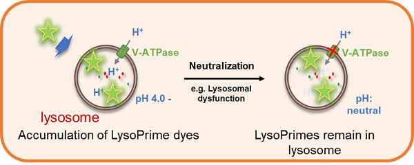
Lysosomal Acidification Inhibitor Included
Fluorescence imaging of lysosomal mass and pH of Bafilomycin A1 (Baf. A1)-induced HeLa cells was performed. The results showed that lysosomal pH was increased in Baf. A1-treated cells compared to control cells, while lysosomal mass was unchanged. Thus, the lysosomal mass and pH was detected at the same time.

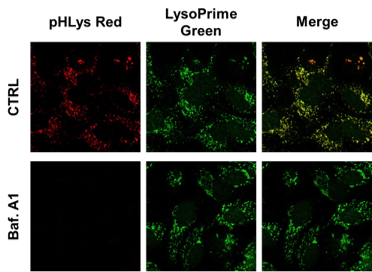
<Experimental Conditions>
LysoPrime Green: Ex = 488 nm, Em = 490 – 550 nm
pHLys Red: Ex = 561 nm, Em = 560 – 620 nm
Q & A
-
Q
Is this right that the transportation temperature and storage temperature (-80°C) are different?
-
A
No problem. There is almost no deterioration during transportation at room temperature. The storage condition is set at -80°C because the purity of the lysosomes was degraded when stored at temperatures other than -80°C for more than one month. We have confirmed that storage at room temperature (25°C) for one month does not affect the staining performance of lysosomes.
-
Q
When I add the stimulation, Is there any precautions?
-
A
Please notice that the LysoPrime Green staining should be performed before the drug stimulation. And the Staining of pHLys Red should be performed after LysoPrime Green staining.
For stimulation with Bafilomycin A1 (lysosomal acidification inhibitor), which is included in the kit, either add Bafilomycin A1 to pHLys Red working solution or add Bafilomycin A1 after the stain with pHLys Red working solution is OK.
-
Q
Can I use medium with serum to staining the cells?
-
A
LysoPrime Green is affected by serum, please prepare the working solution with serum-free medium or HBSS.
On the other hand, pHLys Red can be prepared by medium with serum. Also, you can use HBSS or PBS to prepare it.
-
Q
Can I observe with a fluorescent microscope?
-
A
It is possible to observe with a fluorescent microscope. However, for some cell lines, it may become difficult. In such cases, please use a confocal microscope.
-
Q
What is the recommended final concentration of LysoPrime Green and pHLys Red?
-
A
If you want to optimize the staining conditions, please refer to the following range.
・LysoPrime Green
< When fluorescence intensity is weak >
Please consider using a dilution of 1,000~2,000x.
< If the background fluorescence is observed >
Please consider using a dilution of 2,000~4,000x.
・pHLys Red
< When fluorescence intensity is weak >
Please consider using a dilution of 250~500x.
-
Q
It seems like LysoPrime Green stains other organelles besides lysosomes.
-
A
We have confirmed that the presence of excessive amounts of LysoPrime Green causes the staining of nuclei and cytoplasm in addition to lysosomes. The optimal staining concentration is also affected by cell type and density, so be sure to check the optimal concentration before using the product for the first time. We have confirmed that working solution diluted 2,000-4,000 times with HBSS for 30 minutes can stain lysosomes without any problem.
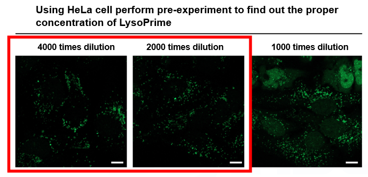
*Scale bar: 10 µm
-
Q
Can I add LysoPrime Green and pHLys Red in the same working solution?
-
A
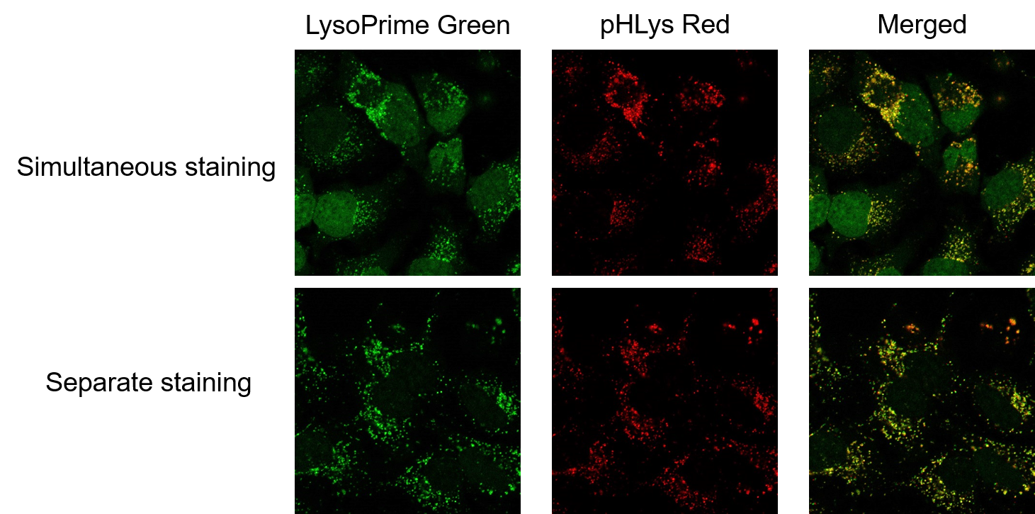
No. Simultaneous staining with LysoPrime Green and pHLys Red changes the localization of LysoPrime Green from Lysosome to other organelles (mainly nucleus and cytosol). That's because pHLys Red causes competitive inhibition with LysoPrime Green in lysosomal localization. Please note that the Staining of pHLys Red must be performed after LysoPrime Green staining.

Handling and storage condition
| Storage Conditions: Freezing (-80℃) Moisture absorption attention | |
|
Danger / harmful symbol mark |

|
|---|---|


