 Hidden sections will not be printed.
General Information
The lysosome is an organelle in which an acid vacuole is formed by a biomembrane. Lysosomes contain various degrading enzymes and contribute to maintaining intracellular homeostasis by acting as a waste disposal system. Recent findings reveal that lysosomal dysfunction is related to some neurodegenerative disorders. Consequently, the investigation of lysosomal function is attracting considerable interest in the scientific community.
Many types of small fluorescent probes are used for monitoring lysosomes in living cells. Dojindo’s LysoPrime Deep Red overcomes known problems with fluorescent lysosome probes, such as lack of specificity for lysosomes and staining dependent on the lysosomal pH. In addition, the high-retentivity of LysoPrime Deep Red enables long-term imaging experiments. And LysoPrime Deep Red emits 633 nm, allowing multiple imaging.
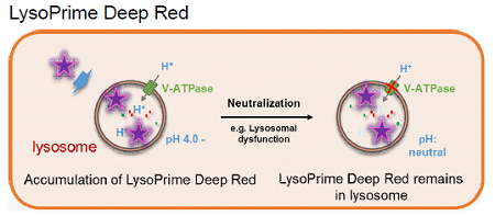
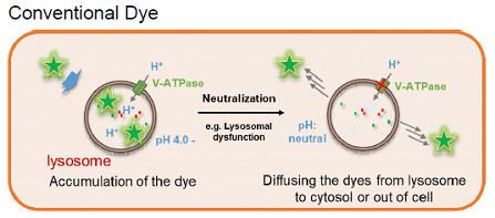
Figure 1. The difference between LysoPrime Deep Red and conventional lysosome staining dye
Fluorescent Property
LysoPrime Deep Redの蛍光特性
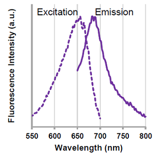 |
λex: 656 nm λem: 682 nm <Recommended filter settings> Ex : 633 nm, Em : 640 – 700 nm |
Contents
| LysoPrime Deep Red | x 1 (corresponds to 10 dishes (35 mm) ) x 3 (corresponds to 30 dishes (35 mm) ) |
Storage Condition
Store at -20 °C and protect from light.
Required Equipment and Materials
- Growth medium
- Hanks’ Balanced Salt Solution (HBSS) or serum-free medium
- Micropipettes
- Microtubes
Preparation of Solution
Preparation of LysoPrime Deep Red DMSO stock solution
Add 20 μl of DMSO to the provided tube containing LysoPrime Deep Red, and dissolve by vortex mixer.
- Store the reconstituted LysoPrime Deep Red DMSO stock solution at −20°C until use.
The solution is stable at −20°C for 1 month.
Preparation of LysoPrime Deep Red working solution
Dilute the LysoPrime Deep Red solution 1,000 times with HBSS or serum-free medium to prepare LysoPrime Deep Red working solution.
- *The final concentration of LysoPrime Deep Red should be optimized depending on the cell line
(dilution range: 1,000 – 5,000 times). - Use HBSS or serum-free medium for the dilution because serum in medium interferes with LysoPrime Deep Red.
- LysoPrime Deep Red working solution should be used on the day it is prepared.
General Protocol
- Seed cells in a dish and culture them overnight at 37 °C in an incubator equilibrated with 95% air and 5% CO2.
- Discard the culture medium and wash the cells twice with HBSS or serum-free medium.
- Add LysoPrime Deep Red working solution to the dish containing the cells and incubate them for 30 minutes at 37 °C in an incubator equilibrated with 95% air and 5% CO2.
- Discard the supernatant and wash the cells twice with HBSS or serum-free medium.
- Add growth medium to the dish, then observe the stained cells under a fluorescence microscope.
Usage Example
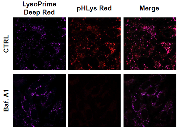
Observation of the lysosomal pH change by confocal fluorescence microscope
- HeLa cells were seeded (1.0 × 10⁴ cells/well) on a μ-slide 8 well plate (ibidi) and cultured overnight at 37 °C in an incubator equilibrated with 95% air and 5% CO2.
- After washing twice with HBSS, 200 μl of LysoPrime Deep Red working solution (1,000 times dilution) and the cells were incubated at 37 °C for 30 min.
- The supernatant was discarded, and the cells were washed twice with HBSS.
- Two hundred (200) μl of pHLys Red (Dojindo code: L265) working solution (HBSS, 1,000 times dilution) containing Bafilomycin A1 (Baf. A1), an inhibitor of lysosomal acidification, was added to the plate, and the cells were incubated at 37 °C for 30 min.
- The supernatant was discarded, and the cells were washed twice with HBSS.
- MEM (200 μl, containing 10% fetal bovine serum) was added to the well, and the stained cells were observed under a confocal fluorescence microscope.
|
Figure 2. The effect of Bafilomycin A1 on lysosomal pH CTRL: Normal condition, Baf. A1: Inhibition of lysosomal acidification (50 nmol/l) |
Frequently Asked Questions / Reference


L264: LysoPrime Deep Red - High Specificity and pH Resistance
Revised Mar., 21, 2023