

|
Recent research on senescence is revealing that senescence is related to lipid droplet and lysosomal dysfunction, which impair cell function. Here are some of the papers showing that these processes leads to neurodegenerative diseases. Cellular senescence, a state of irreversible growth arrest, is closely linked to neurodegeneration through the accumulation of damaged cells in the nervous system. Lipid droplets, which store excess lipids, can accumulate in aging or stressed cells, contributing to cellular dysfunction and exacerbating neurodegenerative processes. Lysosomal dysfunction plays a central role in both lipid accumulation and the removal of cellular waste, and its impairment leads to the accumulation of toxic substances that further drive neurodegeneration. Together, these mechanisms create a cycle of cellular damage that accelerates the progression of neurodegenerative diseases such as Alzheimer's and Parkinson's. |
|||
|
|||
|
|||
|
|||
| Related Applications | |||
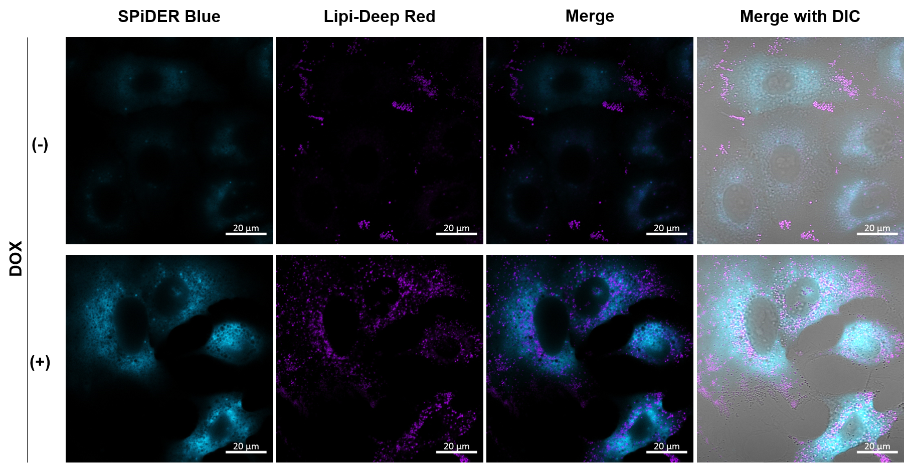
Co-staining with Lipid droplet and SA-β-Gal in fixed cellsImaging analysis of lipid droplet accumulation in senescent cells was performed using normal A549 cells (CTRL) and cells induced senescence by Doxorubicin treatment (DOX). SA-β-Gal was detected as a senescence marker with Cellular Senescence Detection Kit - SPiDER Blue, and lipid droplets were detected with Lipi-Deep Red. As a result, the signal of Lipi-Deep Red was increased in SA-β-Gal-positive senescent cells.
[Detection conditions]
1. A549 (2 x 10⁴) cells were seeded onto µ-slide 8 well plates (ibidi) and cultured overnight in a 37°C CO2 incubator. |
|||
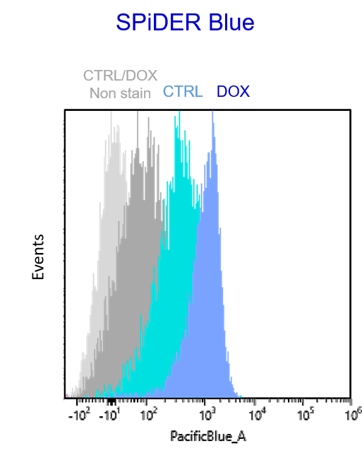
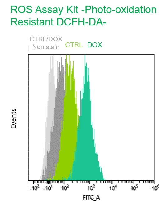
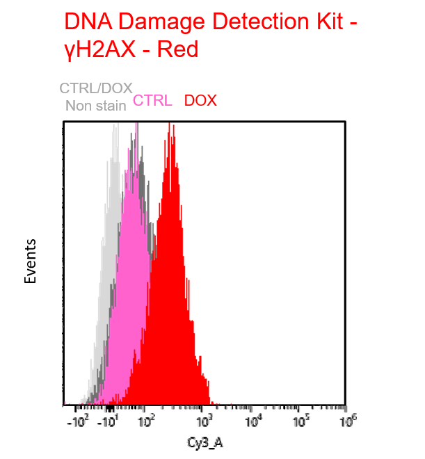
Multiple staining with oxidative stress-related markers using Doxorubicin-induced senescent cells(flow cytometry)Using A549 cells induced to senescence by doxorubicin (DOX) and normal cells (CTRL), changes in oxidative stress-related markers in senescent cells were analyzed by flow cytometry with multiple staining. SA-βGal as a senescence marker was detected by Cellular Senescence Detection Kit - SPiDER Blue, total ROS as an oxidative stress marker was detected by ROS Assay Kit - Photo-oxidation Resistant DCFH-DA-, and γH2AX as a DNA damage marker was detected by DNA Damage Detection Kit - γH2AX-Red. As a result, total ROS and γH2AX were increased in SA-βGal-positive senescent cells, and the increase in oxidative stress-related markers associated with cellular senescence could be detected by multiple staining. <Experimental Procedure> |
|||
| Related Techniques | |||
| Cellular senescence detection | SPiDER-βGal for live-cell imaging or flow cytometry / microplate reader / tissue samples SPiDER-βGal Blue for fixed cell and for multiple staining with immunostaining and other methods |
||
| Lipid Droplet detection | Lipid Droplet Assay Kit - Blue / Deep Red | ||
| Lipid Droplet Staining | Lipi-Blue/ Green/ Red/ Deep Red | ||
| Lysosomal function | Lysosomal Acidic pH Detection Kit-Green/Red and Green/Deep Red | ||
| First-time autophagy research | Autophagic Flux Assay Kit | ||
| Mitochondrial membrane potential detection | JC-1 MitoMP Detection Kit, MT-1 MitoMP Detection Kit | ||
| Mitochondrial superoxide detection | MitoBright ROS Deep Red - Mitochondrial Superoxide Detection | ||
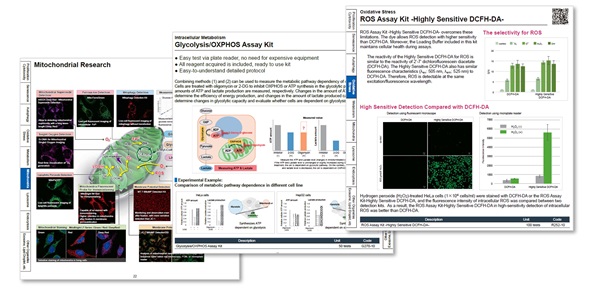
| Glycolysis/Oxidative phosphorylation Assay | Glycolysis/OXPHOS Assay Kit | ||
| Glutathione Quantification | GSSG/GSH Quantification Kit | ||
Senescence and Lipid Droplet Accumulation are Involved in the Onset of Neurodegenerative Diseases
Digital Brochure and Other Information
Curious about other Dojindo products? Discover more with our digital brochure! : Cell Function Analysis
 |
 |
Stay informed and connect with us on our LinkedIn page!
![]()


