


Lysosomal Acidic pH Detection Kit-Green/Deep Red
Lysosomal Acidic pH Detection Kit-Green/Deep Red
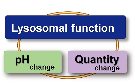
- Accurate pH changes can be detected by measuring lysosomal pH and volume changes in the same sample
- All-in-one Kit with Lysosome Acidification Inhibitor included
- Compatible with imaging and quantitative assay
-
Product codeL268 Lysosomal Acidic pH Detection Kit-Green/Deep Red
| Unit size | Price | Item Code |
|---|---|---|
| 1 set | Find your distributors | L268-10 |


| 1 set | LysoPrime Deep Red pHLys Green Bafilomycin A1 |
x 1 x 1 x 1 |
|---|
Description
Lysosomes are acidic vesicles, one of the intracellular organelles that contribute to the maintenance of homeostasis by degrading unwanted substances. Since lysosomal dysfunction is deeply involved in the onset and progression of neurodegenerative diseases and other disorders, detailed analysis of lysosomes has attracted much attention in elucidating pathological conditions and developing therapeutic agents. Furthermore, recent studies have shown that a decrease in acidity inside lysosomes in mouse models of the neurodegenerative disease Alzheimer's disease results in the inability to degrade waste products inside autophagosomes and the accumulation of toxic amyloid-β*, thus increasing the need to confirm the pH of lysosomes.
Live imaging using small molecule fluorescent dyes has been widely used for lysosomal live cell analysis, but the specificity of the dyes and the accuracy of fluorescence brightness in response to changes in pH have been cited as issues. This product is bundled with two types of dyes that overcome these issues, and the combination of these dyes enables a more accurate live cell analysis of lysosomes.
*Nature Neuroscience, 2022, 25, 688–701.
Lysosomal Analysis Products
| Product Name | Lysosomal pH Detection Dyes and Fluorescence Properties |
Lysosomal Quantity Detection Dyes and Fluorescence Properties |
|---|---|---|
| Lysosomal Acidic pH Detection Kit | pHLys Red Ex: 561 nm / Em: 560-650 nm |
LysoPrime Green Ex: 488 nm / Em: 500-600 nm |
| Lysosomal Acidic pH Detection Kit - Green/Deep Red | pHLys Green Ex: 488 nm / Em: 490-550 nm |
LysoPrime Deep Red Ex: 633 nm / Em: 640-700 nm |
| pHLys Red - Lysosomal Acidic pH Detection | pHLys Red Ex: 561 nm / Em: 560-650 nm |
|
| LysoPrime Deep Red - High Specificity and pH Resistance | LysoPrime Deep Red Ex: 633 nm / Em: 640-700 nm |
|
| LysoPrime Green- High Specificity and pH Resistance | LysoPrime Green Ex: 488 nm / Em: 500-600 nm |


Manual
Technical info
With existing reagents, it was difficult to determine whether lysosomal mass or their function (pH) fluctuated because the discussion was based on changes in the fluorescence brightness of a single dye. This kit contains pHLys Green, which is highly specific to lysosomes and shows pH-dependent changes in fluorescence, and pH-resistant LysoPrime Deep Red [Code:L264]. Using these two dyes, lysosomal pH and volume of the same sample can be measured for a detailed analysis of lysosomal function.
Comparative Performance Analysis
Compared to existing staining dyes, Dojindo's lysosomal detection dyes selectively accumulate on lysosomes and continue to stain for more than 24 hours. These dyes can be used in combination, and lysosomal mass and pH can be more accurately confirmed.
All data were generated in-house under identical imaging and treatment conditions.
Lysosome tracking dye
| Dye | LysoPrime Green | LysoPrime Deep Red | Lysosomal Tracker Green Major commercial supplier |
|
pH Stability |
Resistant to pH elevation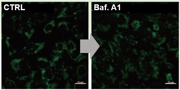 |
Resistant to pH elevation |
Reduced under pH elevation |
|
Lysosomal Retention Time |
Long-term Retention Stable Localization (>24 h) 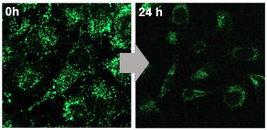 |
Long-term Retention Stable Localization (>24 h) 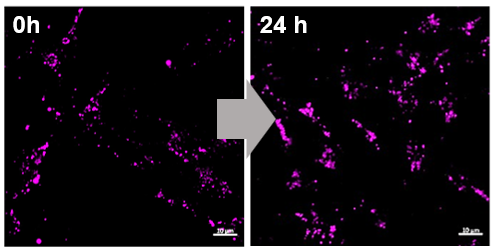 |
Rapid Signal Decline <90 min (identical conditions) |
Lysosomal pH Sensors
| Dye | pHLys Green | pHLys Red | Lysosomal pH Sensor Green Major commercial supplier |
|
pH Stability |
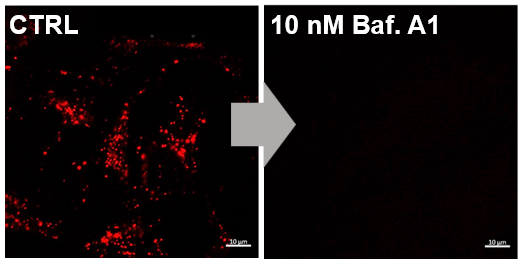
High pH Responsiveness Signal clearly reduced under Baf. treatment 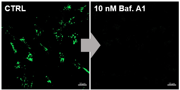 |
High pH Responsiveness |
Less sensitive |
|
Lysosomal Retention Time |
Long-term Retention Stable Localization (>24 h) 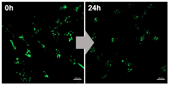 |
Long-term Retention Stable Localization (>24 h) 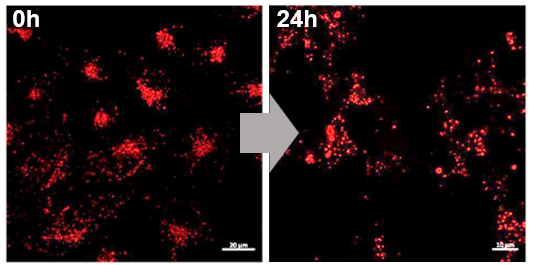 |
Rapid Signal Decline <90 min (identical conditions) 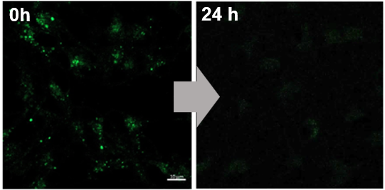 |
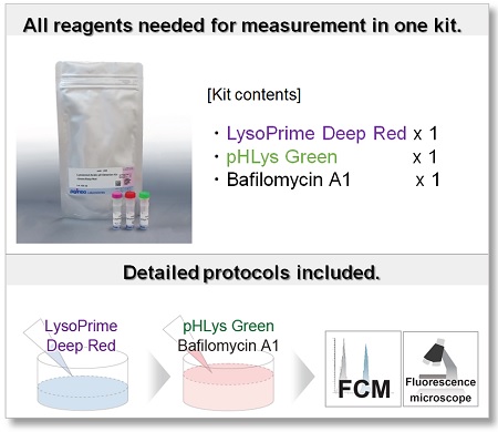
Features of the kit and comparison with existing reagents
All reagents necessary for the measurement are included in one kit. In addition, the fluorescent properties of the dyes enable co-staining with dyes that have red fluorescent properties.
Compared to existing staining dyes, Dojindo's lysosomal detection dyes selectively accumulate on lysosomes and continue to stain for more than 24 hours. These dyes can be used in combination, and lysosomal mass and pH can be more accurately confirmed.
|
【this product】 |
Company T | Company T | ||||
|---|---|---|---|---|---|---|
| Dye/ Wave length |
pHLys Green Ex=488 nm |
Ex=633 nm |
Ex=561 nm |
Ex=488 nm |
Lysosomes pH sensor |
Lysosomes |
| Purpose |
pH |
Mass |
pH |
Mass |
pH |
pH/Mass |
| lysosome pH sensitivity |
✓ |
Resistant to pH change |
✓ |
Resistant to pH change |
Less sensitivity | Less sensitivity |
| lysosome Specificity |
✓ |
✓ |
✓ |
✓ |
✓ |
✓ |
| lysosome retention |
✓ |
✓ |
✓ |
✓ |
✓ |
✓ |
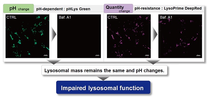
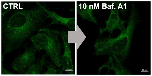
Example of experiment : Imaging and FCM analysis of lysosomal pH change using Bafilomycin A1
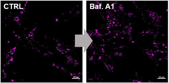
HeLa cells were used to detect changes in lysosomal mass and pH when treated with the lysosomal acidification inhibitor Bafilomycin A1 (Baf. A1). The fluorescence of LysoPrime Deep Red has no change regardless of the addition of Baf. A1, while the fluorescence of pHLys Green decreased due to lysosomal neutralization caused by the addition of Baf. A1. This indicates that Baf. A1 does not affect lysosomal mass, although lysosomal function is reduced.
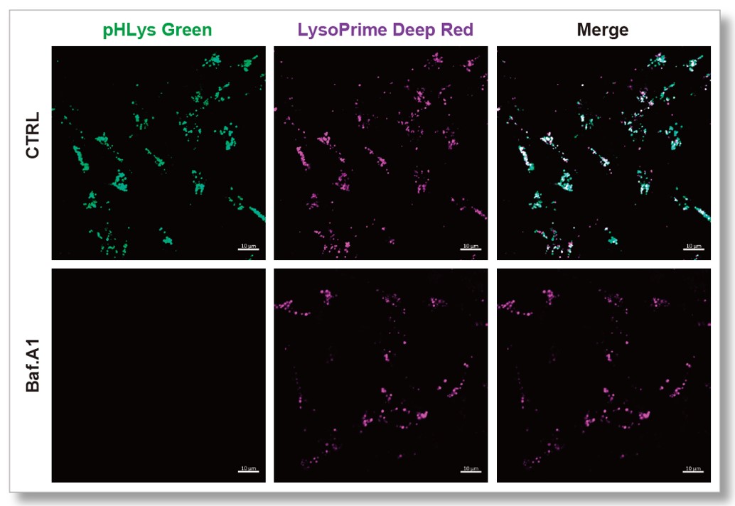 pHLys Green (Green) : Ex=488 nm, Em=490-550 nm LysoPrime Deep Red (Violet) : Ex=633 nm, Em=640-700 nm |
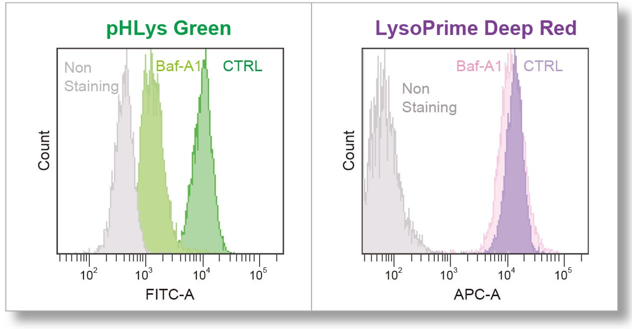 pHLys Green (FITC Filter) : Ex=488 nm, Em=515-545 nm LysoPrime Deep Red (APC Filter) : Ex=640 nm, Em=650-670 nm |
[Protocol for Flow cytometry]
1. HeLa cells (5x10⁴ cells/ml) were seeded (1.0 x 10⁵ cells/well, 2 ml) on 6 well plate and cultured overnight.
2. After washing twice with HBSS, 2 ml of LysoPrime Deep Red working solution (HBSS, 800 times dilution) and the cells were incubated at 37℃ for 30 min.
3. The supernatant was discarded, and the cells were washed twice with HBSS.
4. Two ml of pHLys Green working solution (HBSS, 1,000 times dilution) was added to the plate, and the cells were incubated at 37℃ for 30 min.
5. The supernatant was discarded, and the cells were washed twice with HBSS.
5. Cells were treated with trypsin.
6. Cells were collected and washed with HBSS.
7. The fluorescence of the cells was detected by Flow cytometer.
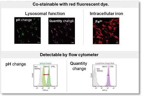
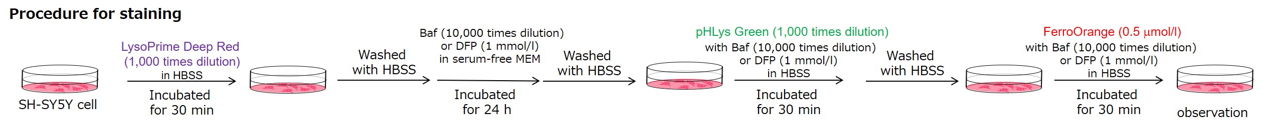
Experimental example: measurement of intracellular iron changes and lysosomal pH changes
In neurodegenerative diseases, the relationship between lysosomal function and iron has attracted attention, and it has been reported* that lysosomal neutralization prevents the breakdown of iron stores (Transferrin or Ferritin), resulting in a decrease in intracellular iron.
Lysosomal pH changes and intracellular iron changes in the same sample were detected using SH-SY5Y cells supplemented with lysosomal acidification inhibitor (Bafilomycin A1) or iron chelator (Deferipron (DFP)). (Lysosomal pH: this kit, Intracellular iron: FerroOrange [Code:F374])
The results showed that the addition of Bafilomycin A1 decreased the fluorescence of FerroOrange, confirming the decrease in intracellular iron. The fluorescence of LysoPrime DeepRed remained almost unchanged, while the fluorescence of pHLys Green decreased due to lysosomal neutralization. These results suggest that there is a relationship between changes in intracellular iron and lysosome function.
*Mol Cell., 2020, 77(3), 645-655.
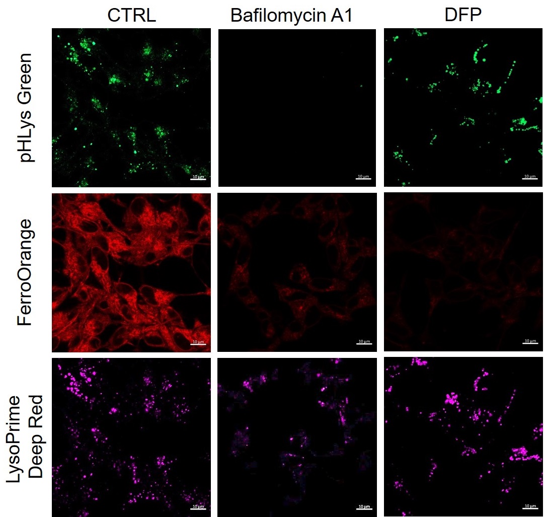
<Condition>
pHLys Green (Green) : Ex=488 nm, Em=486-574 nm
FerroOrange (Red) : Ex=561 nm, Em=550-650 nm
LysoPrime Deep Red (Violet) : Ex=633 nm, Em=599-700 nm
Lysosome Staining Reagents and Kits
Explore Dojindo's wide range of lysosomal staining and pH detection dyes. Choose the following kit or reagent that aligns with your experimental requirements.
| Product Name (Item Code) |
Supported Devices | Indicator and Detection Color | Dyes and Fluorescence Properties |
Approximate Number of Use |
||
|---|---|---|---|---|---|---|
 |
 |
 |
||||
| Lysosomal Acidic pH Detection Kit-Green/Deep Red (L268) | ✓ | ✓ | ✓ | pH | pHLys Green Ex: 488 nm / Em: 490-550 nm |
[for 1 set] 35 mm dish: 10 dishes μ-Slide 8 well: 10 plates 96-well Plate: 2 plates |
| quantity | LysoPrime Deep Red Ex: 633 nm / Em: 640-700 nm |
|||||
| Lysosomal Acidic pH Detection Kit (L266) | ✓ | Need G/Y Laser G:532 nm Y:561 nm |
✓ | pH | pHLys Red Ex: 561 nm / Em: 560-650 nm |
|
| quantity | LysoPrime Green Ex: 488 nm / Em: 500-600 nm |
|||||
| pHLys Red- Lysosomal Acidic pH Detection (L265) | ✓ | ✓ | pH | pHLys Red Ex: 561 nm / Em: 560-650 nm |
[for 1 tube] 35 mm dish: 10 dishes μ-Slide 8 well: 10 plates 96-well Plate: 2 plates |
|
| LysoPrime Deep Red - High Specificity and pH Resistance (L264) | ✓ | ✓ | ✓ | quantity | LysoPrime Deep Red Ex: 633 nm / Em: 640-700 nm |
|
| LysoPrime Green- High Specificity and pH Resistance (L261) | ✓ | ✓ | ✓ | quantity | LysoPrime Green Ex: 488 nm / Em: 500-600 nm |
[for 10 μl] 35 mm dish: 10 dishes μ-Slide 8 well: 10 plates 96-well Plate: 2 plates |
References
| No. | Cell Type | Inducer | Instrument | Reference (Link) |
|---|---|---|---|---|
| 1) | Human astrocytes | HIV-1 Tat, Mutant Tat | Microscope | N. Rezagholizadeh, G. Datta, W.A. Hasler, E.C. Nguon, E.V. Smokey, N. Khan, X. Chen, "SLC38A9 is directly involved in Tat-induced endolysosome dysfunction and senescence in astrocytes", Life Sci Alliance, 2025, doi:10.26508/lsa.202503231 |
| 2) | Human astrocytes | TLR7 agonist R837, Tat protein | Microscope | N. Rezagholizadeh, G. Datta, W.A. Hasler, E.C. Nguon, E.V. Smokey, X. Chen, "TLR7 Mediates HIV-1 Tat-Induced Cellular Senescence in Human Astrocytes", Aging Cell, 2025, doi:10.1111/acel.70086 |
Q & A
-
Q
I am considering a drug stimulus that affects lysosomal pH. What precautions should we take?
-
A
LysoPrime Deep Red staining must be performed before drug stimulation. Staining with pHLys Green must be done after LysoPrime Deep Red staining. For stimulation with Bafilomycin A1 (lysosomal acidification inhibitor), which is included in the kit, either add Bafilomycin A1 to pHLys Green working solution and perform staining and stimulation at the same time, or stain with pHLys Green working solution and then use Bafilomycin A1 for stimulation. Bafilomycin A1 should be added to pHLys Green working solution and stimulation should be performed simultaneously.
-
Q
Can I stain with serum-containing medium?
-
A
LysoPrime Deep Red cannot be stained because it is affected by serum. Make sure to use a serum-free solution such as HBSS or serum-free medium to make a working solution. pHLys Green can be used for staining. HBSS or PBS can be used as a working solution.
-
Q
What concentrations of LysoPrime Deep Red and pHLys Green should be used when considering staining conditions?
-
A
Please refer to the following when optimizing staining conditions.
- LysoPrime Deep Red
<When fluorescence intensity is weak
Use a dilution of 500 to 1,000 times.
<When nonspecific adsorption is observed>
Please consider between 1,000x and 5,000x dilution.
- pHLys Green
<When fluorescence intensity is weak
Use a dilution of 250-1,000x.
<When nonspecific adsorption is observed
Please consider between 1,000x and 2,000x dilution.
- LysoPrime Deep Red
-
Q
The fluorescence of LysoPrime Deep Red decreased after drug treatment. How can I improve this?
-
A
We recommend considering a reduction in drug concentration or exposure time. If they do not resolve the issue, we recommend using LysoPrime Green as an alternative.
At a concentration 20 times higher than the recommended concentration of Bafilomycin A1 (1000nM vs 50nM), LysoPrime Green demonstrated higher retention within lysosomes than LysoPrime Deep Red, as it binds directly to lysosomal proteins.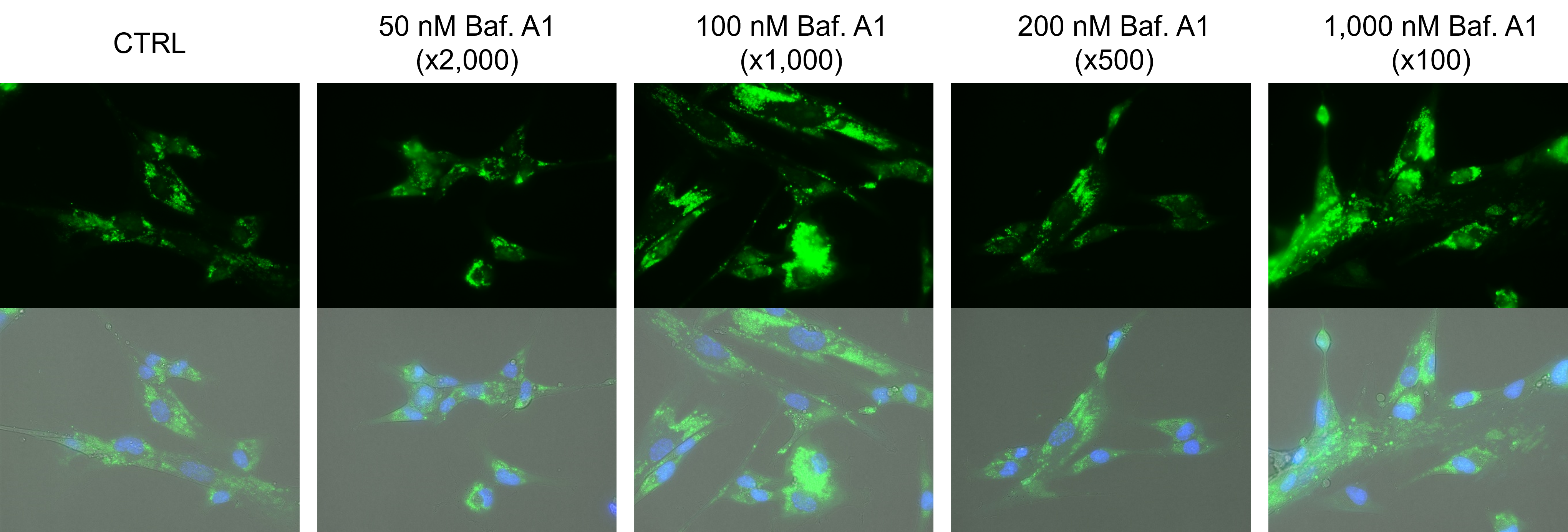
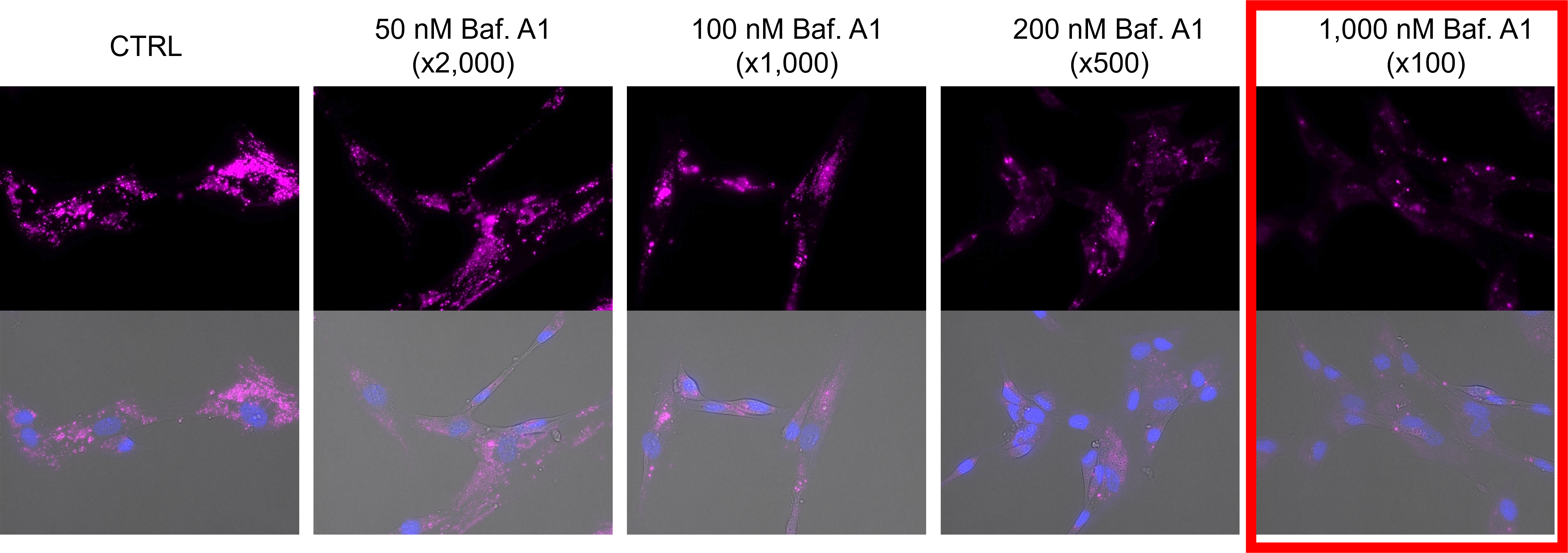
Comparison of LysoPrime Green / Deep Red retention at different bafilomycin A1 concentrations
• LysoPrime Green: recommended concentration x2,000 (recommended concentration range: x1,000 – 4,000)
• LysoPrime Deep Red: recommended concentration x1,000 (recommended concentration range x1,000–5,000)
• Bafilomycin A1: recommended concentration x2,000 (recommended concentration range x1,000–5,000)
-
Q
Can cells be fixed after staining with LysoPrime Deep Red or pHLys Green?
-
A
Fixation is not recommended.
Handling and storage condition
| 1. Storage: -20℃, Protect from light 2. Nitrogen substitution, Moisture-proof |
Related products
The following related products are also used in research related to this product.
-
ROS Detection
ROS Assay Kit -Photo-oxidation Resistant DCFH-DA-
-
Endocytosis Detection
ECGreen-Endocytosis Detection
-
Lysosome Staining Dye Green
LysoPrime Green - High Specificity and pH Resistance
-
Cell Membrane Staining
PlasMem Bright Green
-
Lysosome Staining Deep Red
LysoPrime Deep Red - High Specificity and pH Resistance
-
Lysosomal pH Detection Reagent Red
pHLys Red - Lysosomal Acidic pH Detection


Product Classification
Product Classification
-
Cell Proliferation / Cell Cytotoxicity Assay
Cell Proliferation / Cell Cytotoxicity Assay Kits /Related Reagents
-
Cell Staining
Cell Double Staning Kit /Live Cell Staining /Dead Cell Staining /Nuclear Staining /Mitochondria Staning /Tissue Staining /Nucleolus Staining /Lipid Droplet Staining /Cell Membrane Staining /Lysosome Staining
-
Intracellular Fluorescent Probes
Reagents for Intracellular Calucuum Ion /Reagents for Intracellular Ion /Related Reagents
-
Labeling Chemistry
Protein Labeling Kits /Protein Labeling Reagents /HPLC Derivertization Reagents /Biotion Labeling Reagents /Related Reagents /Exsosome Labeling
-
Oxidative Stress
Stress Maker Detection /NO Detection /NO Donor /NO Inhibitor /ACE Inhibition Assay /Reagents・Kits for Sulfur Biology /Antioxidant Assay Kit /Donors for Sulfur Biology
-
-Bacstain- Series
Bacterial Proliferation Assay Kit /Bacteria Staining /Bacterial Fluorescent Staining
-
Molecular Biology
Transfection Reagents /Nuclear Staining /Agarose /Related Reagents /Buffer for Molecular Biology
-
Detergents
Detergents /Sets
-
Cross-Linking Reagents
Hetero-bifunctional Reagents /Homo-bifunctional Reagents /Others
-
Redox Dyes
Reductive Chromogenic Dyes /Electron Mediators /Oxidative Chromogenic Dyes /Trinder Reagents
-
Ion Analysis
Ionophores /Anion Eliminator /Solvent for Ion Electrode of Liquid Film Type
-
Buffers
Buffers
-
Metal Chelates
EDTA /Other Chelator /Reagents for Chelator Titration
-
Chromogen/Metal Indicator
Chromogen/Metal Indicator
-
Biochemicals
Biochemicals
-
Functional Organic Material
Alkanethiol Derivative /Phosphonic Acid Derivatives
