



Cellular Senescence Detection Kit - SPiDER-βGal
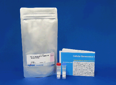
Cellular Senescence Detection
- Fluorescence quantification of SA-β-gal activity
- Compatible with live and fixed cells
- Measured with microscopy or flow cytometry
- One plate is sufficient for one 6-well plate, seven 35mm dishes, or seven chamber slides
-
Product codeSG04 Cellular Senescence Detection Kit - SPiDER-βGal
| Unit size | Price | Item Code |
|---|---|---|
| 1 plate | $281.00 | SG04-01 |
| 3 plates | $620.00 | SG04-03 |
| 10 plates | $1332.00 | SG04-10 |
The same amount of SG03-10 is with SG04-03.
*Difference between SG03 and SG04 is the only amount of reagent.

| 1 plate | ・SPiDER-βGal ・Bafilomycin A1 |
×1 ×1 |
|---|---|---|
| 3 plates | ・SPiDER-βGal ・Bafilomycin A1 |
×3 ×3 |
| 10 plates | ・SPiDER-βGal ・Bafilomycin A1 |
×10 ×10 |
Description
Cellular Senescence, a state of irreversible growth arrest, can be triggered to prevent proliferation of DNA-damaged cells. Increased activity of senescence-associated β-galactosidase (SA-β-gal) is a widely-used marker of cellular senescence. Common SA-β-gal reporters (e.g. X-gal) suffer from incompatibility with live cells, long staining protocols, and poor quantification.
The Cellular Senescence Detection Kit–SPiDER-βGal offers a fast, simple, and quantitative method for SA-β-gal activity detection. The fluorescent reporter, SPiDER-βGal, possesses high cell permeability and high intracellular retention after cleavage by SA-β-gal. Included in this kit is Bafilomycin A1 (BafA1) an inhibitor for endogenous β-galactosidase activity. Pretreatment with BafA1 allows for specific SA-β-gal activity measurement in live cells via flow cytometry or fluorescence microscopy.
Cellular Senescence Analysis Products



Manual


Technical info
SPiDER-βGal Allows for Senescence Detection in Live Cells
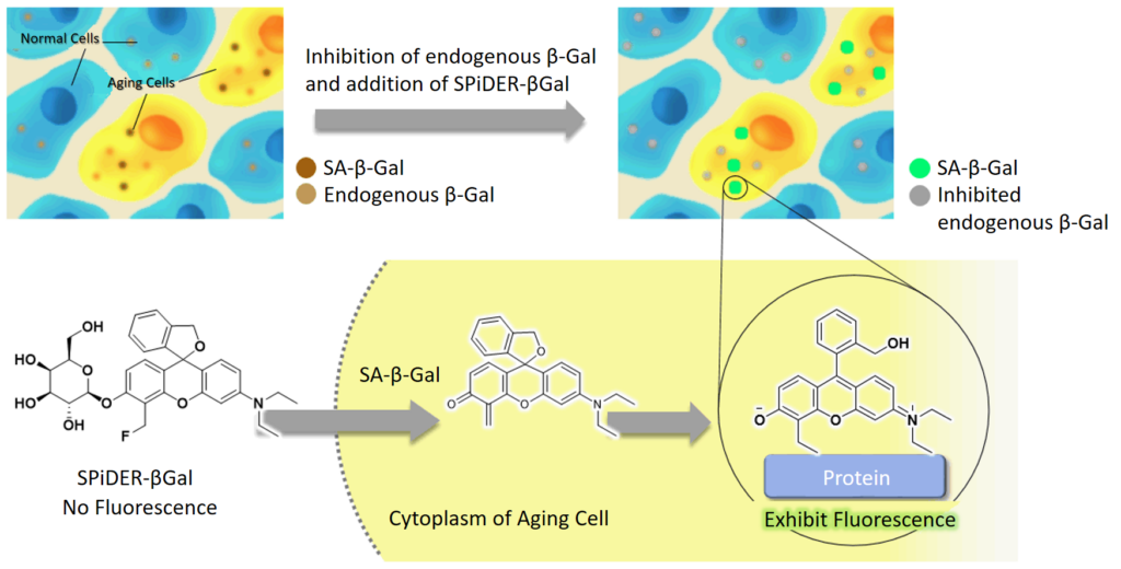
SPiDER-βGal is membrane-permeable and can penetrate live cells without requiring permeabilization or fixation. Within the cell, SPiDER-βGal reacts with SA-β-Gal and forms covalent bonds with neighboring proteins allowing for high retention of SPiDER-βGal. Addition of BafA1 before SPiDER-βGal staining suppresses the activity of endogenous β-galactosidase in live cells, allowing specific detection of SA-β-Gal activity via fluorescence.
SPiDER-βGal Spectrum
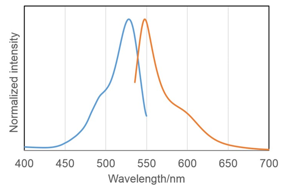
λex:488~540nm, λem: 530~570nm
Quantify Senescence in 30-Minutes

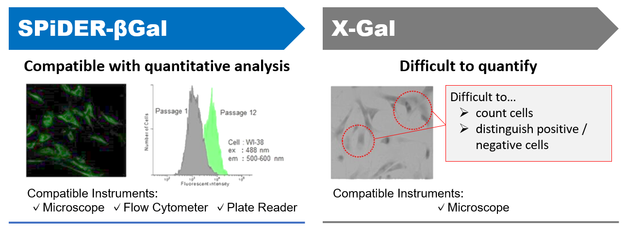
SPiDER-βGal Fluorescence is More Quantitative than X-Gal
SPiDER-βGal Outperforms C12FDG and X-Gal
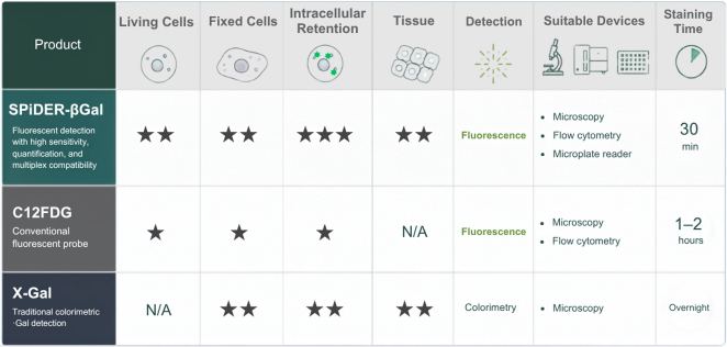
Objective fluorescence-based detection enables quantitative analysis, multiplex staining, and shorter workflows compared with conventional colorimetric staining methods.
Approximate Number of Samples that Can Be Analyzed
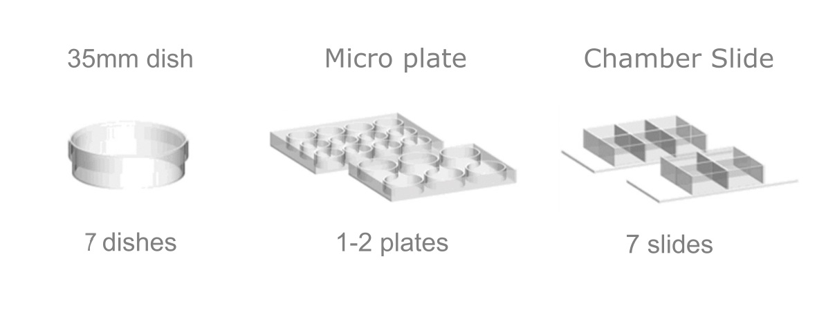
Usage may vary depending on total volume of SPiDER-βGal Working Solution used for staining.
Application and Experimental Data
SA-β-Gal Activity in Live Cells
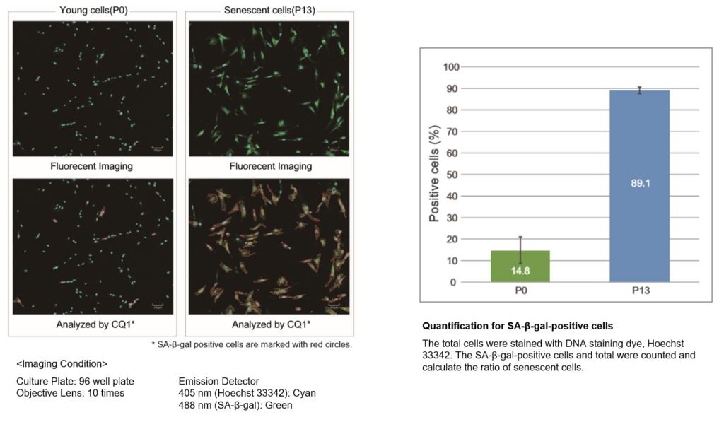
Young (passage = 0) and senescent (passage = 13) WI-38 cells were co-stained with SPiDER-βgal, Hoechst 33342 and visualized with confocal microscopy. SA-β-gal-positive cells (SPiDER-βgal) and total cells (Hoechst 33342) were counted under microscope to calculate the percent of the senescent cells. Stained cells were analyzed using confocal quantitative image cytometer CQ1.
SPiDER-βGal and γ-H2A.X Costaining in Fixed Cells
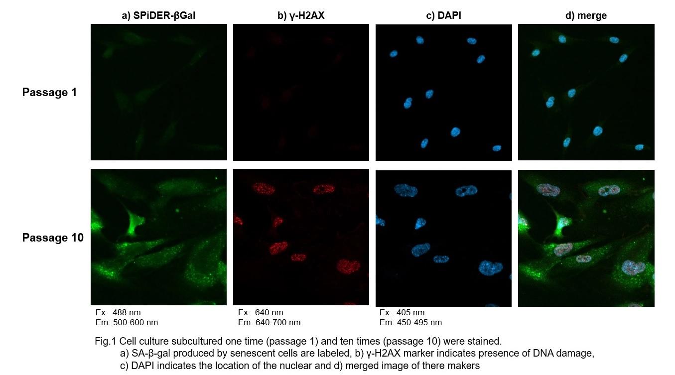
SPiDER-βGal enables co-staining with dyes and immunofluorescence markers. DNA Damager marker γ-H2AX (Alexa Fluor 647, Red) and DNA (DAPI, Blue). This multi-spectral workflow provides deeper biological insight than SA-β-Gal staining alone.
FCM SA-β-Gal Activity Measurement in Suspension Cells
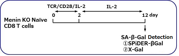
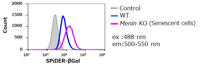
Prof. Masakatsu Yamashita's research group at Ehime University Graduate School of Medicine has shown Menin controls T cell exhaustion, aging, and maintains normal immune function. SPiDER-B-Gal fluorescence was quantified via FCM in WT and Menin-KO cells by stimulating TCR (T cell receptor) in the presence of interleukin 2 (IL-II) in naive CD8+ T cells. *Data were kindly provided by Masakatsu Yamashita.
Quantification of Cell Senescence, Cell Cycle, and Mitochondrial Function
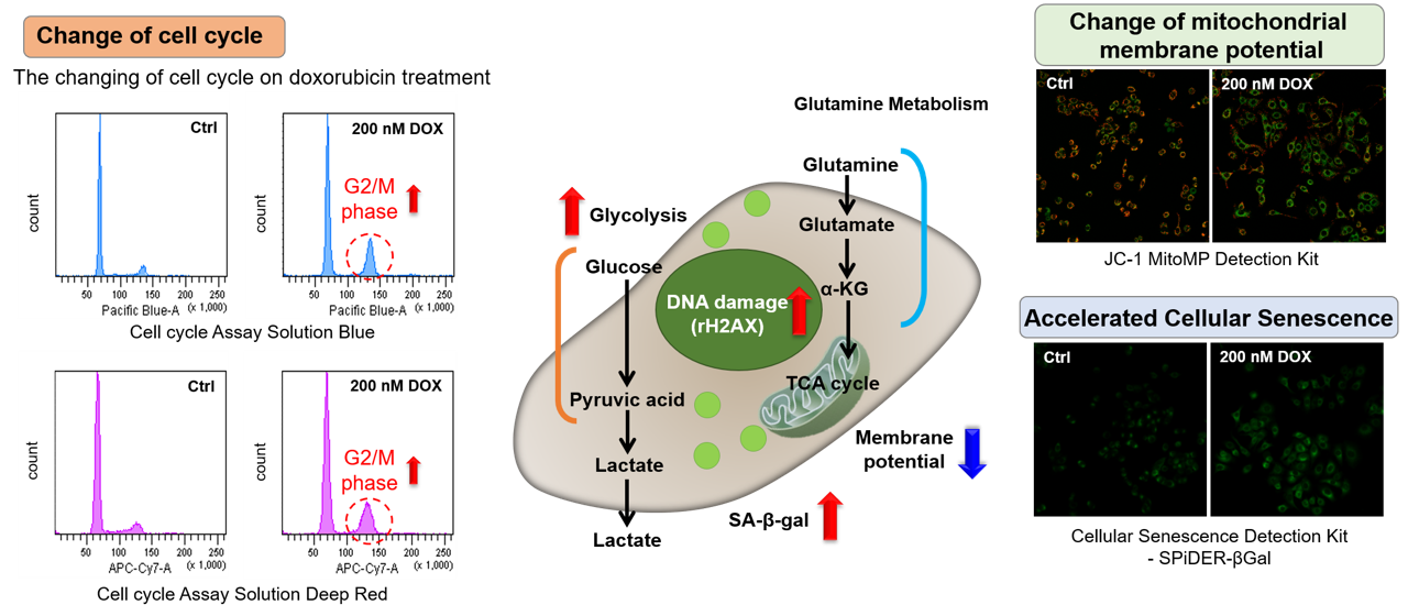
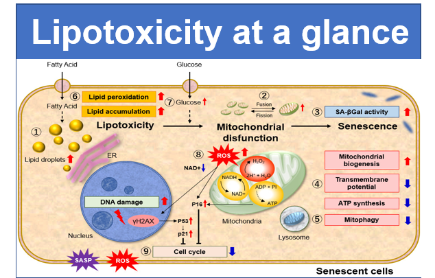
Doxorubicin (DOX) acts to inhibit cell proliferation during G2/M phases of the cell cycle and induces cellular senescence. A549 cells treated with 200nM DOX, showed increased number of cells in G2/M phase (Cell Cycle Assay Solution Blue and Deep Red), induced cellular senescence (SPiDER-βGal), and altered mitochondrial membrane potential (JC-1 MitoMP Detection Kit).
References
Q & A
-
Q
What is the reason of adding Bafilomycin A1 (Baf. A1)?
-
A
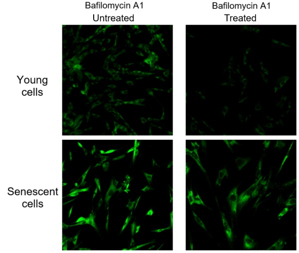
Endogenous β-galactosidase existing in living cells interfere with selective detection of SA-β-Gal. Baf. A1 is an inhibitor of ATPase in lysosome. pH in lysosome is kept neutral by adding Baf. A1. Cellular Senescence Detection Kit - SPiDER-βGal contains Baf. A1 which allows to detect SA-β-Gal selectively. Baf. A1 is utilized for living cell assays only. Baf. A1 is not used in fixed cells because intracellular pH is controlled with the buffer.
The figure above shows the difference in SA-β-Gal detection with and without Baf. A1. Baf. A1 is also known as an autophagy inhibitor. When using Baf. A1. on live cells, please consider whether there are any effects on the experiment and if so, we recommend experiments with fixed cells. Baf. A1 is not used with fixed cells as the intracellular pH is controlled by buffer. Fixed cells are stained with SPiDER-βGal as procedure described in the manual.
-
Q
How stable is DMSO stock solution?
-
A
SPiDER-βGal DMSO stock solution and Bafilomycin A1 DMSO stock solution is stable for 1 month at -20 ℃.
-
Q
How stable is working solution?
-
A
SPiDER-βGal working solution and Bafilomycin A1 working solution can’t be stored. Be sure to use the working solution immediately.
-
Q
Is there any advice for observing senescent cells?
-
A
Lipofuscin is a fluorescent pigment that accumulates in a variety of cell types with age. Lipofuscin consists of autofluorescent granules and may results in high background for fluorescence microscopy. In order to achieve accurate SA-β-gal activity assay in senescent cells, we recommend to prepare samples without SPiDER-βGal staining as a negative control. Please compare fluorescence intensity of both cells with or without SPiDER-βGal staining.
> For Flow Cytometry Detection
Step 1. Prepare senescent cells and non-senescent cells. Measure MFI (Mean Fluorescence Intensity) of samples below.
[Senescent cells]
Sample A: The cells stained with SPiDER-βGal
Sample B: The cells without SPiDER-βGal staining
[Non-senescent cells]
Sample A’: The cells stained with SPiDER-βGal
Sample B’: The cells without SPiDER-βGal staining
Step 2. Calculate SA-β-gal activity (senescent cells) with the following formula
SA-β-gal activity (senescent cells) = MFI of Sample A - MFI of Sample B
Step 3. Calculate SA-β-gal activity (non-senescent cells) with the following formula
SA-β-gal activity (non-senescent cells) = MFI of Sample A’ - MFI of Sample B’
- Determine the SA-β-gal activity by comparing the SA-β-gal activity between senescent cells and non-senescent cells.
- Change of SA-β-gal activity associated with senescence = (Value from Step 2- value from Step 3)
>For Microscopy
Step 1. Prepare senescent cells without SPiDER-βGal staining and observe fluorescent image.
Step 2. Adjust detection sensitivity in microscopy to reduce background autofluorescence of lipofuscin.
Step 3. Observe fluorescent image of senescent cells and non-senescent cells under the settled condition in step 2.
-
Q
Can SPiDER-βGal be used with fixed cells?
-
A
It is possible. If fixed cells are stained with SPiDER-βGal, it is not necessary to pre-treat the cells with bafilomycin A1. However, McIlvain buffer adjusted to pH 6.0 must be prepared separately. See manual for details.
-
Q
What is the protocol for detecting fixede and stained cells using a flow cytometer?
-
A
See protocol below.
(1) Prepare cells in 35 mm dishes and incubate overnight at 37°C in a 5% CO2 incubator.
(2) After removing the culture medium, wash the cells once with 2 ml of HBSS.
(3) Treat the cells with trypsin and mix with 500 µl serum containing medium to collect the cells.
(4) Centrifuge at 300 xg for 5 minutes and remove the supernatant.
(5) Suspend the cells in 100 µl 2% PFA/PBS to the cells and incubate for 5 minutes at room temperature.
(6) Centrifuge at 300 xg for 5 minutes and remove the supernatant.
(7) Suspend the cells in 500 µl HBSS, centrifuge at 300 ×g for 5 minutes and remove the supernatant. Repeat this step twice.
(8) Add 500 µL of SPiDER-βGal working solution (for fixed cells) and incubate for 30 minutes at 37°C.
Note: Do not use a 5% CO2 incubator due to reduce pH fluctuations.
(9) Centrifuge at 300 xg for 5 minutes and remove the supernatant.
(10) Suspend the cells in 500 µl HBSS, centrifuge at 300 ×g for 5 minutes and remove the supernatant. Repeat this step twice.
(11) Suspend the cells in 500 µl HBSS and analyze on a flow cytometer.
-
Q
Can SPiDER-βGal staining and immunostaining be performed after fixation of the cells?
-
A
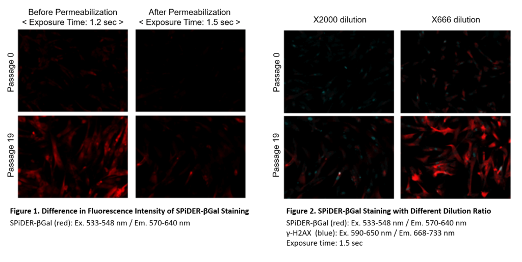
Preparation of SPiDER-βGal working solution
Dilute the SPiDER-βGal DMSO stock solution 2,000 times *1 with McIlvaine buffer (pH 6.0).
*1 Fixation and permeablization could leads to lower sensitivity (Figure 1), if you need higher signals,dilute the SPiDER-βGal DMSO stock solution 500 – 1,000 times with the McIlvaine buffer (Figure 2).Preparation of McIlvaine buffer (pH 6.0)
Mix 0.1 mol/l citric acid solution (3.7 ml) and 0.2 mol/l sodium phosphate solution (6.3 ml). Confirm the pH is 6.0. If the pH is not 6.0, adjust the pH by adding either citric acid solution or sodium phosphate solution. Dilute this buffer 5 times with ultrapure water.Staining procedure (35 mm dish)
1. Prepare cells on 35 mm dish for assay and culture the dish at 37℃ overnight in a 5% CO2 incubator.
2. Remove the culture medium. Add 2 ml of 4% paraformaldehyde (PFA) /PBS solution to the cells and incubate at room temperature for 3 minutes *2.
*2 Avoid a longer treatment period, which leads to decrease in SA-β-gal activity.
3. Remove the supernatant, and wash the cells 3 times with 2 ml of PBS.
4. Add 2 ml of SPiDER-βGal working solution and incubate at 37℃ for 30 minutes*3.
*3 We recommend not to use a 5% CO2 incubator for fixed cell experiments. If incubation is done in a 5% CO2 incubator, the pH of the buffer may become acidic. Acidic pH results in higher background from the endogenous β-galactosidase activity and it would be difficult to distinguish between normal cells and senescent cells.
5. After removing the supernatant, wash the cells twice with PBS.
6. Add 0.1% Triton X-100/PBS to cells and incubate for 30 minutes at room temperature.
7. Wash the cells twice with PBS.
8. Add 1% BSA/PBS to the cells and incubate for 1 hour at the room temperature
9. Add anti- γ-H2AX antibody (mouse) diluted with 1% BSA/PBS to the cells and incubate at 4℃ overnight.
10. Wash the cells 3 times with PBS.
11. Add anti- mouse secondary antibody (Cy5) diluted with 1% BSA/PBS to the cells and incubate at room temperature for 1 hour.
12. Wash cells twice with PBS and observe under a fluorescence microscope.
-
Q
Is there any advice if the fluorescence of stained cells is low?
-
A
Please check the following points.
(1) Use the filter that matches the fluorescence characteristics of the reagent.
<Recommended filters>
Fluorescence microscope: excitation (500-540 nm), fluorescence (530-570 nm)
Flow cytometer: excitation (488 nm), fluorescence (500-540 nm)
(2) Use fresh working solution(3) Increase the staining time.
If fluorescence cannot be confirmed after 30 minutes of incubation after addition of SPiDER-βGal working solution, increase the incubation time to 45-60 minutes.
-
Q
Can the cells be fixed after staining with SPiDER-βGal?
-
A
It is possible. Fixation with 4% paraformaldehyde is recommended.
-
Q
Does serum or phenol red in the medium affect detection?
-
A
Serum and phenol red in the medium do not affect the detection of SA-β-gal.
-
Q
No difference in fluorescence intensity between senescent cells and control cells
-
A
Cellular senescence wasn’t induced. Please prepare for positive control. Please visit product page and check “Positive Control”
-
Q
Background in fix cells assay
-
A
The incubation with SPiDER-βGal working solution was done in a 5% CO2 incubator. We recommend not to use a 5% CO2 incubator during incubation with SPiDER-βGal working solution. If incubation is done in a 5% CO2 incubator, the pH of the buffer may become acidic. Acidic pH results in higher background from the endogenous β-galactosidase activity and it would be difficult to distinguish between control cells and senescent cells. Please incubate the plate in a dry incubator without CO2 .
Handling and storage condition
| 0-5°C |

Related products
The following related products are also used in research related to this product.


Product Classification
Product Classification
-
Cell Proliferation / Cell Cytotoxicity Assay
Cell Proliferation / Cell Cytotoxicity Assay Kits /Related Reagents
-
Cell Staining
Cell Double Staning Kit /Live Cell Staining /Dead Cell Staining /Nuclear Staining /Mitochondria Staning /Tissue Staining /Nucleolus Staining /Lipid Droplet Staining /Cell Membrane Staining /Lysosome Staining
-
Intracellular Fluorescent Probes
Reagents for Intracellular Calucuum Ion /Reagents for Intracellular Ion /Related Reagents
-
Labeling Chemistry
Protein Labeling Kits /Protein Labeling Reagents /HPLC Derivertization Reagents /Biotion Labeling Reagents /Related Reagents /Exsosome Labeling
-
Oxidative Stress
Stress Maker Detection /NO Detection /NO Donor /NO Inhibitor /ACE Inhibition Assay /Reagents・Kits for Sulfur Biology /Antioxidant Assay Kit /Donors for Sulfur Biology
-
-Bacstain- Series
Bacterial Proliferation Assay Kit /Bacteria Staining /Bacterial Fluorescent Staining
-
Molecular Biology
Transfection Reagents /Nuclear Staining /Agarose /Related Reagents /Buffer for Molecular Biology
-
Detergents
Detergents /Sets
-
Cross-Linking Reagents
Hetero-bifunctional Reagents /Homo-bifunctional Reagents /Others
-
Redox Dyes
Reductive Chromogenic Dyes /Electron Mediators /Oxidative Chromogenic Dyes /Trinder Reagents
-
Ion Analysis
Ionophores /Anion Eliminator /Solvent for Ion Electrode of Liquid Film Type
-
Buffers
Buffers
-
Metal Chelates
EDTA /Other Chelator /Reagents for Chelator Titration
-
Chromogen/Metal Indicator
Chromogen/Metal Indicator
-
Biochemicals
Biochemicals
-
Functional Organic Material
Alkanethiol Derivative /Phosphonic Acid Derivatives
