|
Ferroptosis suppressor protein-1 (FSP1) has recently been identified as a second system that suppresses ferroptosis, preventing lipid peroxidation independently of the glutathione-GPX4 axis. Researchers have used a small molecule library screen to discover 3-phenylquinazolinones, represented by icFSP1, as potent inhibitors of FSP1. Unlike previous inhibitors, icFSP1 does not directly inhibit FSP1 enzyme activity, but causes FSP1 to relocate from the membrane and condense, working in synergy with GPX4 inhibition.
Learn more about how the authors detected Lipid peroxide, one of the ferroptosis markers, using Liperfluo. We also offer ferroptosis-related products: FerroOrange, Mito-FerroGreen.
|
|
-
Phase separation of FSP1 promotes ferroptosis
Click here for the original article: T. Nakamura, et. al., Nature (2023)
Point of Interest
- Ferroptosis suppressor protein-1 (FSP1) suppresses ferroptosis, preventing lipid peroxidation independently of the glutathione-GPX4 axis.
- Using a small molecule library screen, 3-phenylquinazolinones, represented as icFSP1, are potent inhibitors of FSP1.
- icFSP1 does not directly inhibit FSP1 enzyme activity, but causes FSP1 to relocate from the membrane and condense, working in synergy with GPX4 inhibition.
- icFSP1 has been found to reduce tumor growth and induce FSP1 condensates in tumors in vivo
|
|
Related Techniques
|
- Intracellular / mitochondrial ferrous ion (Fe2+) detection
- FerroOrange(intracellular), Mito-FerroGreen(mitochondrial)
|
- NAD(H) and NADP(H) redox couples assay
- NAD/NADH and NADP/NADPH Assay Kit
|
|
|
- Lipid Peroxidation Assay
- Lipid Peroxidation Probe -BDP 581/591 C11-
|
- Lipid droplets detection
- Lipi-Blue / Green / Red / Deep Red
|
- Total ROS detection
- Highly sensitive DCFH-DA or Photo-oxidation Resistant DCFH-DA
|
- Glutathione Quantification
- GSSG/GSH Quantification Kit
|
- Cystine Uptake detection
- Cystine Uptake Assay Kit
|
- MDA detection
- MDA Assay Kit
|
|
Related Applications
|
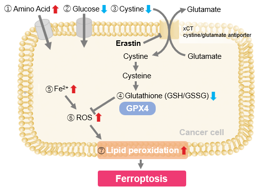
Erastin-Induced Ferroptosis: Evaluating Intracellular Uptake and Redox Balance
We investigated the transition of cellular metabolisms in A549 cells treated with erastin, a known ferroptosis inducer. Our results revealed the following.

- The inhibition of cystine uptake by erastin led to a depletion of cysteine, which in turn increased the compensatory uptake of other amino acids.
- Glucose uptake, which typically promotes ferroptosis*, was found to decrease upon erastin treatment, suggesting a potential cellular self-defense mechanism.
- The depletion of cysteine resulted in a decrease in glutathione levels and an increase in Fe2+, ROS, and lipid peroxides, all of which are recognized markers of ferroptosis.
Cell Line: A549
Incubation Conditions: 100 μmol/l Erastin/MEM, 37℃, 3h
*Reference: Xinxin Song, et al., Cell Reports, (2021)
Products in Use
|


