


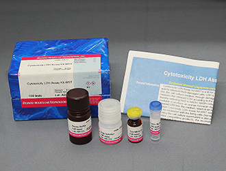
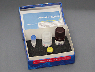
Cytotoxicity LDH Assay Kit-WST
Cytotoxicity Assay
- Can be used with and without transferring supernatant
- Stable Working Solution (6 months at 0-5 oc)
- No need to freeze the Working Solution
-
Product codeCK12 Cytotoxicity LDH Assay Kit-WST
| Unit size | Price | Item Code |
|---|---|---|
| 100 tests | Find your distributors | CK12-01 |
| 500 tests | Find your distributors | CK12-05 |
| 2000 tests | Find your distributors | CK12-20 |


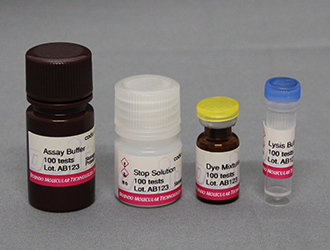
| 100 tests | ・Dye Mixture ・Assay Buffer ・Lysis Buffer ・Stop Solution |
×1 11 ml×1 1.1 ml×1 5.5 ml×1 |
|---|---|---|
| 500 tests | ・Dye Mixture ・Assay Buffer ・Lysis Buffer ・Stop Solution |
×1 55 ml×1 5.5 ml×1 27.5 ml×1 |
| 2000 tests | ・Dye Mixture ・Assay Buffer ・Lysis Buffer ・Stop Solution |
×4 55 ml×4 5.5 ml×4 27.5 ml×4 |
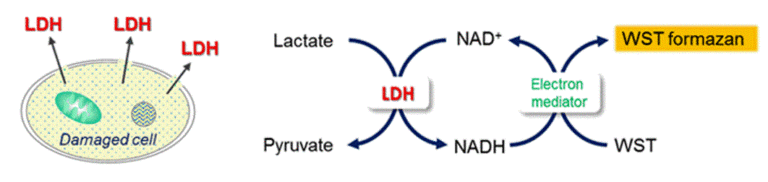
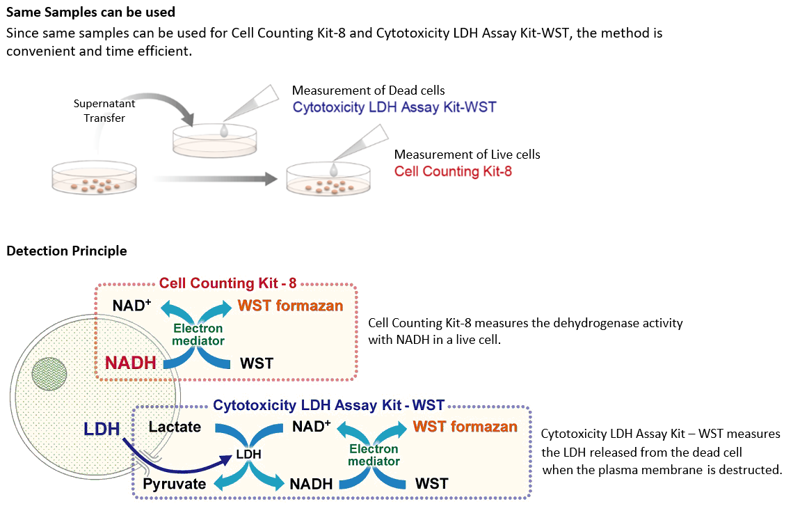
Detection Principle
Lactate dehydrogenase(LDH) is an enzyme that presents in almost cell types and it catalyzes the oxidation of lactate to pyruvate in the presence of co-enzyme NAD+. Once cells are impaired by stress, injuries, chemicals, or intercellular signals, LDH is rapidly released from the cell membrane. Thus, the measurement of the amount of released LDH from cells is one of the major methods to assess the cell death. Since Dojindo’s Cytotoxicity LDH Assay Kit-WST neither reflects the activity of living cells nor is harmful to cells, it allows the assay to perform in wells containing both viable and damaged cells.
| Developer | Dojindo Molecular Technologies, Inc. |
|---|


Manual
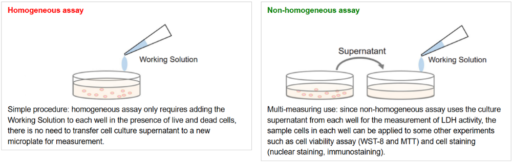
Homogeneous Assay
Non-homogeneous Assay
Optimization of cell concentration
Technical info
Flexible Application Methods
Cytotoxicity LDH Assay Kit-WST can be used with or without transferring the supernatant. Choose the method that best fits your experiment.
In assays with suspension cells, the method without supernatant transfer eliminates the need for a microplate centrifuge to settle cells at the bottom of the plate.
Video Guide
How to Use the Homogeneous Assay(without transfer) and Non-homogeneous Assay (with transfer)
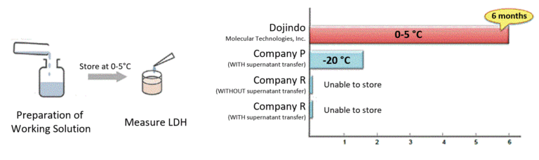
Stable Working Solution
Working Solution is stable for 6 months under refrigerated conditions. Therefore, after the preparation, Working Solution can be used as a ready-to-use solution at any time during this period.
Nearly 90% lower total cost* compared with short-stability LDH kits.
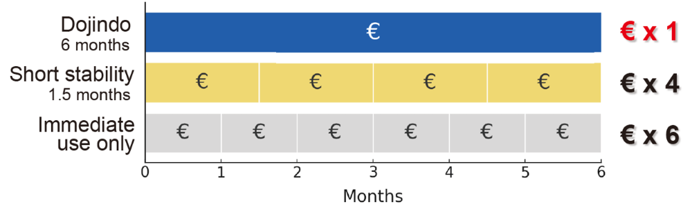
*Based on a comparison of 2000 tests over a continuous six month usage period. Actual results may vary depending on assay frequency and local pricing.
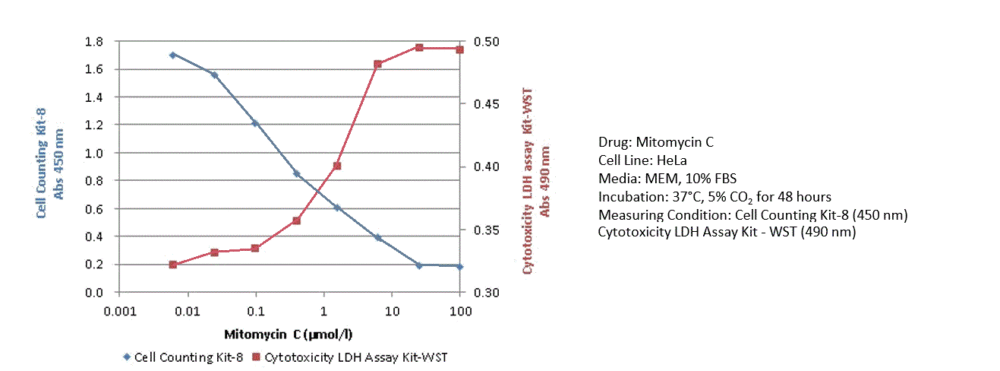
Cytotoxicity of Mitomycin C using HeLa cells
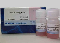
The cytotoxicity of Mitomycin C on HeLa cells was assessed using the Cell Counting Kit-8 and the Cytotoxicity LDH Assay Kit-WST. The Cell Counting Kit-8 reflects intracellular metabolic activity, while the Cytotoxicity LDH Assay Kit-WST detects LDH released from damaged cell membranes. Because the assays measure different indicators, the Mitomycin C concentration at which cytotoxicity was observed varied between the two methods.
Complement-Dependent Cytotoxicity Assay Using RAJI Cells
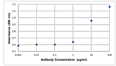
Complement-dependent cytotoxicity (CDC) was evaluated using RAJI cells. An increase in absorbance was observed in a concentration-dependent manner with the anti-CD20 antibody, indicating that binding of the antibody to the cells activated the complement system and resulted in cell lysis.
For reference, see the ADCC Assay manual (a related assay method).
Cell Death Related Products
| Type | Product Name | Target | Detection Properties |
|---|---|---|---|
| Cell viability | Cell Counting Kit-8 | Dehydrogenase Activity |
Plate reader Colorimetric, λ= 450nm |
| Necrosis (Cytotoxicity) |
Cytotoxicity LDH Assay Kit-WST | Leaked LDH | Plate reader Colorimetric, λ= 490nm |
| Apoptosis | Annexin V Apoptosis Plate Assay Kit | Phosphatidylserine | Plate reader Ex: 488 nm / Em: 525 nm |
| Ferroptosis | FerroOrange | Intracellular Fe2+ | Microscopy, FCM, Plate reader Ex: 543 nm / Em: 580 nm |
| Ferroptosis | Mito-FerroGreen |
Mitochondrial Fe2+ |
Microscopy Ex: 505 nm / Em: 535 nm |
| Ferroptosis | Liperfluo | Lipid Peroxide | Microscopy, FCM Ex: 488 nm / Em: 500-550 nm |
References
| Reference No. | Sample | Citation |
|---|---|---|
| 1) | C3H10T1/2 | S. F. Jin, H. L. Ma, Z. L. Liu, S. T. Fu, C. P Zhang, Y. He, "XL413, a cell division cycle 7 kinase inhibitor enhanced the anti-fibrotic effect of pirfenidone on TGF-β1-stimulated C3H10T1/2 cells via Smad2/4.", Exp Cell Res., 2015, 339, (2), 289. |
| 2) | HepG2 | S. Watanabe, C. S. Moniaga, S. Nielsen, M. Hara-Chikuma, "Aquaporin-9 facilitates membrane transport of hydrogen peroxide in mammalian cells.", Biochem Biophys Res Commun ., 2016, 471, 191. |
| 3) | HEECs, Human Esophageal Epithelial Cells | L. Wu, T. Oshima, J. Shan, H. Sei, T. Tomita, Y. Ohda, H. Fukui, J. Watari, H. Miwa , "PAR-2 activation enhances weak acid-induced ATP release through TRPV1 and ASIC sensitization in human esophageal epithelial cells.", Am J Physiol Gastrointest Liver Physiol ., 2015, 309, G695. |
| 4) | Human L02 Hepatocyte | D. Zhou, B-H Li, J. Wang, Y-N Ding, Y. Dong, Y-W Chen and J-G Fan, "Prolyl Oligopeptidase Inhibition Attenuates Steatosis in the L02 Human Liver Cell Line.", PloS ONE., 2016, 11, (10), e0165224. |
| 5) | Human primary Hepatocyte | T. Fukami, A. Iida, K. Konishi, M. Nakajima , "Human arylacetamide deacetylase hydrolyzes ketoconazole to trigger hepatocellular toxicity", Biochem. Pharmacol., 2016, 116, 153. |
| 6) | Mouse embryonic fibroblast: MEF | Y. Takanezawa, R. Nakamura, Y. Sone, S. Uraguchi and M. Kiyono, "Atg5-dependent autophagy plays a protective role against methylmercury-induced cytotoxicity", Toxicol. Lett., 2016, 262, 135. |
| 7) | Neural stem/progenitor cell | M. Tanaka, M. Yoneyama, T. Shiba, T. Yamaguchi, K. Ogita, "Protease-activated receptor-1 negatively regulates proliferation of neural stem/progenitor cells derived from the hippocampal dentate gyrus of the adult mouse ", J Pharmacol Sci., 2016, 131, (3), 162 |
| 8) | Primary cultured cortical neuron cell | S. Wakatsuki, A. Furuno, M. Ohshima, and T. Araki, "Oxidative stress-dependent phosphorylation activates ZNRF1 to induce neuronal/axonal degeneration", J Cell Biol., 2015, 211, (4), 881. |
| 9) | DLD1 and AGS | A. Ishijima, K. Minamihata, S. Yamaguchi, S. Yamahira, R. Ichikawa, E. Kobayashi, M. Iijima, Y. Shibasaki, T. Azuma, T. Nagamune & I. Sakuma, "Selective intracellular vaporisation of antibody-conjugated phase-change nano-droplets in vitro", Scientific Reports ., 2017, DOI:10.1038/srep44077. |
| 10) | U87, HUVEC | N. Li, W. Zhang, M. Khan, L. Lin, JM. Lin, "MoS2-LA-PEI nanocomposite carrier for real-time imaging of ATP metabolism in glioma stem cells co-cultured with endothelial cells on a microfluidic system", Biosens Bioelectron., 2017, 99, (2018), 142. |
| 11) | Tissue (Mouse liver) |
Y. Sakurai, W. Mizumura, M. Murata, T. Hada, S. Yamamoto, K. Ito, K. Iwasaki, T. Katoh, Y. Goto, A. Takagi, M. Kohara, H. Suga, and H. Harashima, "Efficient siRNA delivery by lipid nanoparticles modified with a non-standard macrocyclic peptide for EpCAM-targeting", Mol. Pharm.., 2017,10.1021/acs.molpharmaceut.7b00362. |
| 12) | Human pleural mesothelial cell | K. Hattori, K. Nakadate, A. Morii, T. Noguchi, Y. Ogasawara, K. Ishii., "Exposure to nano-size titanium dioxide causes oxidative damages in human mesothelial cells: The crystal form rather than size of particle contributes to cytotoxicity.", Biochem. Biophys. Res. Commun.., 2017,10.1016/j.bbrc.2017.08.054. |
| 13) | Tissue (murine distal colon) |
H. Sakai, S. Tabata, M. Kimura, S. Yabe, Y. Isa, Y. Kai, F. Sato, T. Yumoto, K. Miyano, M. Narita and Y. Uezono, "Active Ingredients of Hange-shashin-to, Baicalelin and 6-Gingerol, Inhibit 5-Fluorouracil-Induced Upregulation of CXCL1 in the Colon to Attenuate Diarrhea Development", Biol. Pharm.Bull.., 2017, 40, (12), 2134. |
| 14) | SH-SY5Y | R. Watanabe, T. Kurose, Y. Morishige and K. Fujimori, "Protective Effects of Fisetin Against 6-OHDA-Induced Apoptosis by Activation of PI3K-Akt Signaling in Human Neuroblastoma SH-SY5Y Cells.", Neurochem. Res.., 2018, 43, (2), 488-499. |
| 15) | MC3T3-E1[Derived from mouse cranial crown] | A. Oyane, M. Nakamura, I. Sakamaki, Y. Shimizu, S. Miyata and H. Miyaji, "Laser-assisted wet coating of calcium phosphate for surface-functionalization of PEEK", PLoS ONE ., 2018, doi:10.1371/journal.pone.0206524. |
| 16) | CRC Cell [Colorectal cancer], HT29 Cell[Adenocarcinoma of the human colon] | T. Fuji, Y. Umeda, A. Nyuya, F. Taniguchi, T. Kawai, K. Yasui, T. Toshima, K. Yoshida, T. Fujiwara, A. Goel and T.Nagasaka, "Detection of Circulating MicroRNAs with Ago2 Complexes to Monitor the Tumor Dynamics of Colorectal Cancer Patients during Chemotherapy.", Int. J. Cancer., 2018, doi: 10.1002/ijc.31960 . |
| 17) | SFXN2 knockout HEK293 | E. E. Mon, F. Y. Wei, R. N. R. Ahmad, T. Yamamoto, T. Moroishi and K. Tomizawa, "Regulation of mitochondrial iron homeostasis by siderofexin 2 ", J Physiol Sci., 2018, doi:10.1007/s12576-018-0652-2. |
| 18) | Human umbilical vein cell line [EA.Hy926] | Y. Xu, C. Huang, M. Liu, N. Chen, W. Chen, C. Yang, Y. Zhao, X. Li, J. Duan, S. Liu and S. Yang, "Survivin regulated by autophagy mediates hyperglycemia-induced vascular endothelial cell dysfunction", Exp. Cell Res.., 2018, doi: 10.1016/j.yexcr.2018.01.037. |
| 19) | MIAPaCa-2[Human Pancreatic Cancer] | M. Akimoto, R. Maruyama, Y. Kawabata, Y. Tajima and K. Takenaga, "Antidiabetic adiponectin receptor agonist AdipoRon suppresses tumour growth of pancreatic cancer by inducing RIPK1/ERKdependent necroptosis", Cell Death Dis., 2018, 9, 804. |
| 20) | Fibro adipogenic progenitor cells:FAPs | Y. Saito, T. Chikenji, T. Matsumura, M. Nakano and M. Fujimiya, "Exercise enhances skeletal muscle regeneration by promoting senescence in fibro-adipogenic progenitors", Nat. Commun., 2020, doi:10.1038/s41467-020-14734-x. |
| 21) | TIG-3 (human normal fibroblasts), PANC-1 (human pancreatic cancer cell line), MIA PaCa-2 (human pancreatic cancer cell line) | T. M. Loo, X. Zhou, Y. Tanaka, S. Sugawara, S. Yamauchi, H. Kawasaki, Y. Matsuoka, Y. Sugiura, S. Sakuma, Y. Yamanishi, S. Yotsumoto, K. Dodo, Y. Shirasaki, T. Kamatani and A. Takahashi, "Senescence-associated lysosomal dysfunction impairs cystine deprivationinduced lipid peroxidation and ferroptosis", Nat. Commun., 2025, doi.org/10.1038/s41467-025-61894-9 |
| 22) | Calu-1, H661(Lung cancer–derived), HT1080 [fibrosarcoma](Sarcoma) | Y. Saimoto, D. Kusakabe, K. Morimoto, Y. Matsuoka, E. Kozakura, N. Kato, K. Tsunematsu, T. Umeno, T. Kiyotani, S. Matsumoto, M. Tsuji, T. Hirayama, H. Nagasawa, K. Uchida, S. Karasawa, M. Jutanom and K. Yamada, "Lysosomal lipid peroxidation contributes to ferroptosis induction via lysosomal membrane permeabilization", Nat. Commun., 2025, doi.org/10.1038/s41467-025-58909-w |
Q & A
-
Q
Can I measure with filters other than 490 nm?
-
A
We recommend 490 nm, but a difference of 490±2-3 nm is no problem. 450 nm filters can be used as we have experience with them. Please note that the absorbance will be higher than with the 490 nm filter.
-
Q
Is there any correlation with Cell Counting Kit-8 in toxicity testing?
-
A
Cytotoxicity LDH Assay Kit-WST and Cell Counting Kit-8 do not necessarily correlate with each other due to their different measurement principles. Therefore, when testing for cytotoxicity, it is recommended to use several types of indicators, such as the LDH Assay and the Cell Counting Kit-8.
- Cytotoxicity LDH Assay Kit-WST: Measures LDH activity released into the medium due to cell membrane damage (cell membrane damage is an indicator).
- Cell Counting Kit-8: Measures intracellular metabolic activity (dehydrogenase activity is an indicator).
-
Q
The LD50 value differs significantly from previous measurements. Is there anything I can do?
-
A
The LD50 value may be affected by the test substance depending on the state of the cells. The LD50 value is affected by the state of the cells.
The LD50 value may be more reliable if a narrower dilution factor is used when preparing a dilution series of the test substance.
It is recommended that the concentration of the test substance to be used be determined according to the following procedure.<Study procedure>
(1) Repeat the 10-fold dilution based on the highest available concentration of the test substance and perform toxicity testing over a wide sample concentration range of about 5-6 digits. [Figure 1]. Using the operation in (1), make a rough estimate of the highest predicted concentration in the concentration range at which toxicity will occur.
(2) Based on the result of (1), repeat 2-fold dilution based on the predicted maximum concentration (the lowest concentration that causes 100% lethality) at which the concentration of the substance to be tested appears, and perform toxicity testing in a concentration range covering 2-3 orders of magnitude. [Figure 2].
(3) Repeat step (2) to finally establish a concentration range in which at least 3 sample concentrations between 20-80% cytotoxicity can be plotted. [Figure 3]
-
Q
Can you measure different isoenzymes of LDH?
-
A
This product is designed to measure cytotoxicity using total LDH activity as an indicator, so it cannot measure LDH1, LDH2, LDH3, etc. separately.
-
Q
Can I store samples?
-
A
It can be stored for approximately one week in refrigerated storage1).
Storage at room temperature2) or freezing is not recommended due to inactivation of LDH.
1) M. Allen, P. Millett, E. Dawes, N. Rushton., "Lactate dehydrogenase activity as a rapid and sensitive test for the quantification of cell numbers in vitro.", Clin Mater. 1994; 16, (4), 189.
2) A. Wagner, A. Marc, JM. Engasser, A. Einsele, "The use of lactate dehydrogenase (LDH) release kinetics for the evaluation of death and growth of Biotechnol Bioeng. 1992; 39, (3), :320.Stability may vary depending on evaluation conditions and other factors. Please refer to the above paper with understanding.
-
Q
Does the Cytotoxicity LDH Assay Kit-WST include LDH standards?
-
A
No, LDH standards are not included in the kit as the kit is intended for cytotoxicity testing.
-
Q
Is it necessary to test immediately after adding Stop Solution?
-
A
Yes, the assay should be performed within 3 hours after the addition of the Stop Solution. In this case, please protect the plate from light.
-
Q
There is a lot of variation between wells, what could be the cause?
-
A
There is a lot of variation between wells, what could be the cause?
Please check the following possible reasons.
(1) Air bubbles are formed on the surface of the culture medium.
Please remove the air bubbles using the following methods.
Remove air bubbles with the tip of a syringe or syringe needle.
(If you have a 96-well plate centrifuge) Centrifuge at 1,000 x g for 1 minute.(2) The reagent concentration has changed due to evaporation of water from the culture medium during incubation.
The outermost wells of the microplate are prone to water evaporation during incubation. When incubating for a long time, do not use the outermost wells for measurement, but use only the culture medium in them.
(3) When adding reagents, there is a large time difference between the first and last wells to be added.
For Working Solution and Stop Solution, which are added to all wells, the use of a multichannel pipette is recommended.
(4)The added reagents are not well mixed.
When adding Lysis Buffer, Working Solution and Stop Solution, mix them with a plate mixer or gently tap the side of the plate with your finger to mix the added reagents and medium.
Note that the amount of Lysis Buffer added is small and will affect the High Control value.
When tapping the plate, be careful not to spill the medium in the wells.(5) The weight of the (multichannel) pipette used is not accurate.
Please check if the pipette weight is accurate.
-
Q
The background is now high. What is the cause and what can I do about it?
-
A
The following causes are possible.
(1) LDH present in serum in the medium is increasing the background. Use serum-free medium or medium prepared with 5% or less serum.
(2) Strong reducing substances in the test substance or medium may cause the WST dye to miscolorize and increase the background. Confirm this by measuring a reagent blank.
(In addition, common culture media such as DMEM, RPMI, F-12, etc. usually do not contain reducing agents).
-
Q
Small difference in absorbance between high and low controls during cell count optimization.
-
A
(1) The absorbance of the low control may be higher due to high background. Check the following
LDH in serum in the medium increases the background. Use serum-free medium or medium prepared with 5% or less serum.
If the test sample or culture medium contains strong reducing substances, the WST dye will mischromatize and increase the background. Confirm this by measuring a reagent blank.(2) The High Control may contain viable cells.
To ensure that the Lysis Buffer is evenly distributed in the wells and that all cells are dead cells, gently vortex the plate after adding the Lysis Buffer to mix the Lysis Buffer thoroughly.
-
Q
The percent of cytotoxicity is lower than expected, what could be the cause?
-
A
The number of cells after culture in a toxicity test fluctuates due to two factors: not only (1) the decrease in cells caused by the test substance (cytotoxicity), but also (2) the proliferation of cells over time.
When cells are incubated for a long time, such as 24 to 48 hours, the effect of cell proliferation over time (2) is largely reflected.
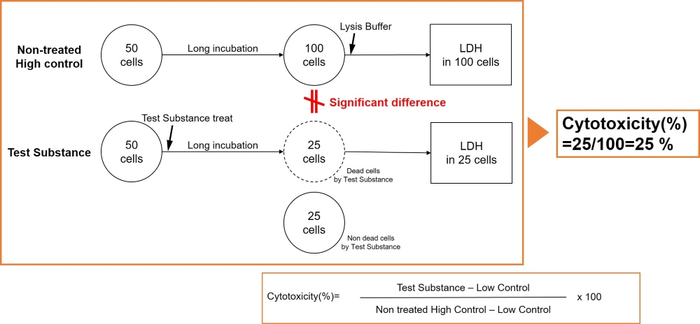
Therefore, in some cases, High control well may not be an appropriate control for the Test substance well because of the large difference in the number of cells between the non-test substance treated High control well and the Test substance well.
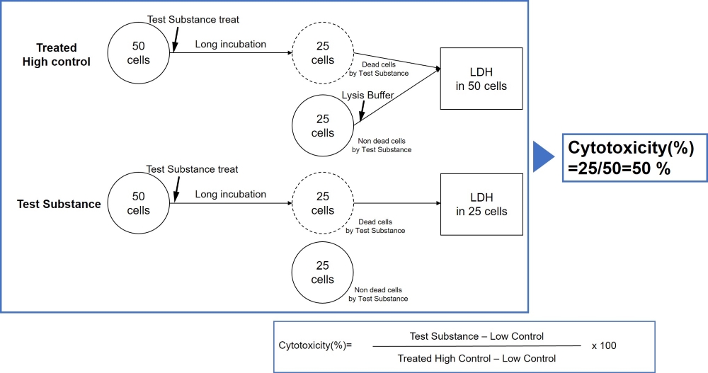
Please prepare a High control with test substance well according to the table below if cells are incubated for more than 24 hours and calculate the percent of cytotoxicity according to the following equation.
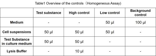
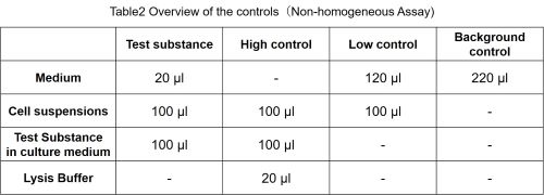
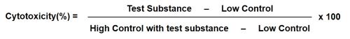
-
Q
Can I purchase the Lysis Buffer separately?
-
A
No, we don’t sell the Lysis Buffer included in the kit separately.
However, if you need additional supplies, you can use the 11% Triton X-100 solution as a substitute.
Handling and storage condition
| 0-5°C, Protect from light | |
|
Danger / harmful symbol mark |
 
|
|---|---|
Related products
The following related products are also used in research related to this product.
-
Cell Proliferation / Cytotoxicity Assay
Cell Counting Kit-8
-
Apoptosis Detection
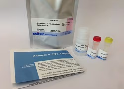
Annexin V, FITC Apoptosis Detection Kit
-
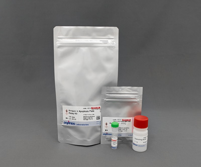
Apoptosis (Annexin V) Plate Assay Kit
Annexin V Apoptosis Plate Assay Kit
-
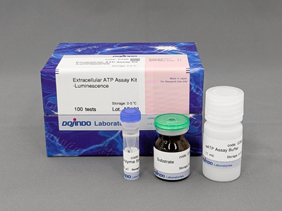
Extracellular ATP Assay Kit-Luminescence
Extracellular ATP Assay Kit-Luminescence
-
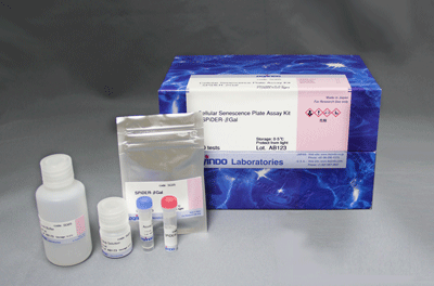
Senescence Cell Detection (for Microplate)
Cellular Senescence Plate Assay Kit - SPiDER-βGal


