


Annexin V, FITC Apoptosis Detection Kit
Apoptosis Detection
-
Product codeAD10 Annexin V, FITC Apoptosis Detection Kit
| Unit size | Price | Item Code |
|---|---|---|
| 50 assays | Find your distributors | AD10-10 |
For Research Use Only Products
| 50 assays | Annexin V, FITC Conjugate PI Solution Annexin V Binding Buffer |
50 assays x 1 bottle 50 assays x 1 bottle 50 assays x 1 bottle |
|---|
Description
Apoptosis is one of the programmed cell death, that plays an important role in maintaining the homeostasis and developmental processes in both plants and animals. Any abnormal cells during the cytogenesis are eliminated by apoptosis. For instance, tumor growth from cancer cells occurred in vivo is inhibited by induction of apoptosis. However, apoptosis is not induced when the error occurs in tumor suppressor gene p53. Thus, the growth of cancer cells has been found to proceed. Apoptotic cells can be identified based on the alteration of cellular morphology as well as the alternation of biomedical changes.
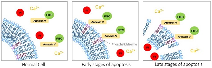
As of today, various indicators have been established such as caspase activity variation, DNA fragmentation and phosphatidylserine transition on the cell surface-chromosomal. Annexin V stained cells are used to indicate cell membrane changes that occur in the early stage of apoptosis. Once apoptosis is initiated, the phosphatidylserine presents in the inner cell membrane migrates through the cell membrane of the lipid bilayer. Annexin V specifically binds to phosphatidylserine in the presence of protein-dependent Ca ion. By using fluorescent-labeled Annexin V, the apoptotic cells can be identified by flow cytometry or fluorescence microscopy.


Manual
Technical info
Experimental Data
|
|
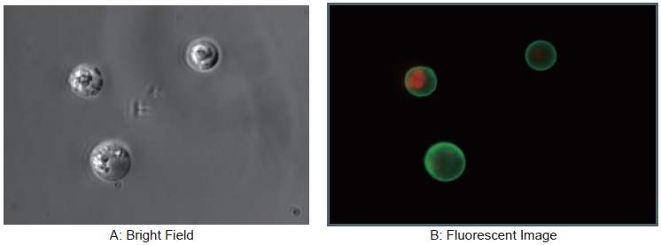
Jurkat cells were apoptosis induced with staurosporine with its concentration of 1 μg/ml at 37°C for 3.5 hours and then observed under a fluorescent microscope.
|
|
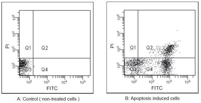
Jurkat cells were apoptosis induced with staurosporine with its concentration of 1μg/ml at 37°C for 3.5 hours and then analyzed with a flow cytometer.
Cell Death Related Products
| Type | Product Name | Target | Detection Properties |
|---|---|---|---|
| Cell viability | Cell Counting Kit-8 | Dehydrogenase Activity |
Plate reader Colorimetric, λ= 450nm |
| Necrosis (Cytotoxicity) |
Cytotoxicity LDH Assay Kit-WST | Leaked LDH | Plate reader Colorimetric, λ= 490nm |
| Apoptosis | Annexin V Apoptosis Plate Assay Kit | Phosphatidylserine | Plate reader Ex: 488 nm / Em: 525 nm |
| Ferroptosis | FerroOrange | Intracellular Fe2+ | Microscopy, FCM, Plate reader Ex: 543 nm / Em: 580 nm |
| Ferroptosis | Mito-FerroGreen | Mitochondrial Fe2+ |
Microscopy Ex: 505 nm / Em: 535 nm |
| Ferroptosis | Liperfluo | Lipid Peroxide | Microscopy, FCM Ex: 488 nm / Em: 500-550 nm |
References
1) Anti-mitotic chemotherapeutics promote apoptosis through TL1A-activated death receptor 3 in cancer cells. Cell Research, 2018,28,544 – 555
2) Nanoenzyme-Augmented Cancer Sonodynamic Therapy by Catalytic Tumor Oxygenation ACS Nano,2018, 12, 3780?3795
3) Bacteria-Driven Hypoxia Targeting for Combined Biotherapy and Photothermal Therapy ACS Nano,2018,12 (6), pp 5995–6005
4) Tumor-Specific Multiple Stimuli-Activated Dendrimeric Nanoassemblies with Metabolic Blockade Surmount Chemotherapy Resistance. ACS Nano,2017,11(1),416-429.
5) Near Infrared-Guided Smart Nanocarriers for MicroRNA-Controlled Release of Doxorubicin/siRNA with Intracellular ATP as Fuel. ACS Nano,2016,10(3),3637-47
6) Noninvasive small-animal imaging of galectin-1 upregulation for predicting tumor resistance to radiotherapy. Biomaterials, 2018,158,1-9
7) Cytotoxicity of guanine-based degradation products contributes to the antiproliferative activity of guanine-rich oligonucleotides. Chemical Science,2015,6,3834-3836
8) A Novel Strategy through Combining iRGD Peptide with Tumor-Microenvironment-Responsive and Multistage Nanoparticles for Deep Tumor Penetration. ACS Appl.Mater.Interfaces, 2015,7 (49),27458–27466
9) Trifolium Repens Blocks Proliferation in Chronic Myelogenous Leukemia via the BCR-ABL/STAT5 Pathway. Cells, 2020, 9 (2), 379
Handling and storage condition
| 0-5oC |
Related products
The following related products are also used in research related to this product.
-
Cell Proliferation / Cytotoxicity Assay
Cell Counting Kit-8
-
Cytotoxicity Assay
Cytotoxicity LDH Assay Kit-WST
-
Apoptosis (Annexin V) Plate Assay Kit
Annexin V Apoptosis Plate Assay Kit
-
Senescence Cell Detection (for Microplate)
Cellular Senescence Plate Assay Kit - SPiDER-βGal
-
ROS Detection
ROS Assay Kit -Highly Sensitive DCFH-DA-


