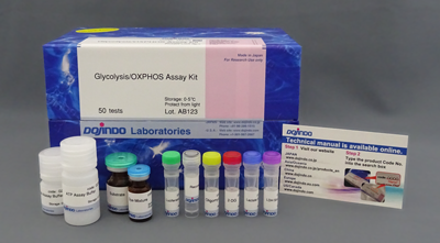
ATP Assay Kit-Luminescence
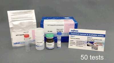
ATP Measurement
- High-sensitivity detection can be performed with stable luminescence.
- Measurement of unstable ATP is not necessary because ATP standard is supplied with the ATP Assay Kit-Luminescence.
- The procedure is very simple, and only required to add the reagent.
-
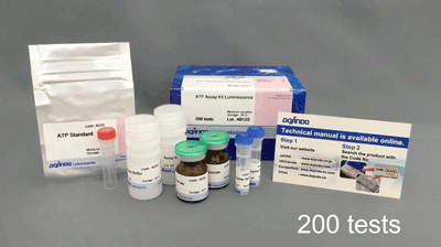
Product codeA550 ATP Assay Kit-Luminescence
| Unit size | Price | Item Code |
|---|---|---|
| 50 tests | Find your distributors | A550-10 |
| 200 tests | Find your distributors | A550-12 |
| 50 tests | ・Enzyme Solution ・Substrate ・Assay Buffer ・ATP Standard |
10 µl×1 ×1 5.5 ml×1 ×1 |
|---|---|---|
| 200 tests | ・Enzyme Solution ・Substrate ・Assay Buffer ・ATP Standard |
20 µl×2 ×2 11 ml×2 ×1 |
Description
Adenosine triphosphate (ATP) is an energy source for living cells and is synthesized by both glycolysis and mitochondrial oxidative phosphorylation. Mitochondria generate 95% of cellular ATP, and mitochondrial dysfunction reduces ATP levels in cells. Therefore, the measurement of ATP levels has been established as a proxy for mitochondrial activity. Decreased ATP levels are associated with cancer, aging, neurodegenerative diseases, and mitochondrial diseases. Cancer cells rely on glycolysis, a process that is less efficient than oxidative phosphorylation to synthesize ATP. However, recent studies have revealed that ATP synthesis in cancer cells shifts from glycolysis to oxidative phosphorylation when glycolysis is suppressed.
The ATP Assay Kit-Luminescence enables the quantitation of intracellular ATP by luciferase luminescence assay. This kit can be applied for microplate assay. Furthermore, this kit can be applied for microplate assay.
Manual
Technical info
The ATP Assay Kit-Luminescence enables the quantitation of intracellular ATP by luciferase luminescence assay. This kit can be applied for microplate assay. Furthermore, this kit can be applied for microplate assay.

Generate a stable luminescent signal with high reproducibility
Without complicated steps such as medium removal and cell washing, the ATP Assay Kit-Luminescence can generate a stable luminescent signal with a half-life of greater than 3 hours by simply mixing the kit components and adding the reagent. This kit contains an ATP Standard, which provides highly reproducible data without measuring unstable ATP.
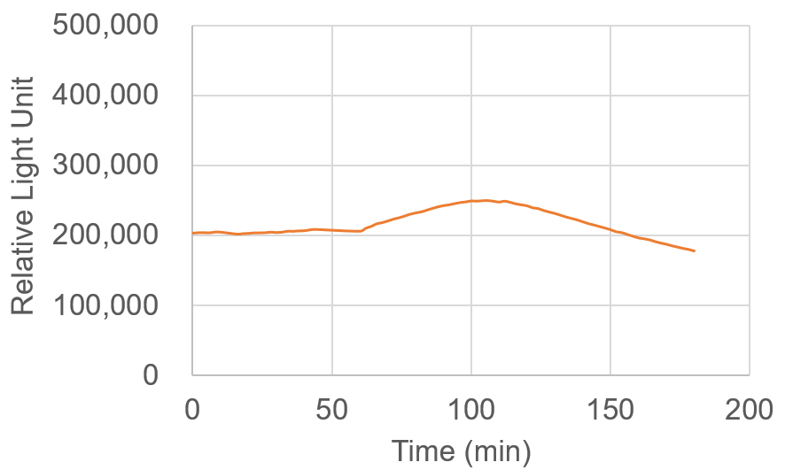
Time-course measurements of relative light units collected 3 hours after the addition of the reagent.
Cell type:HeLa cells (10,000cells/well)
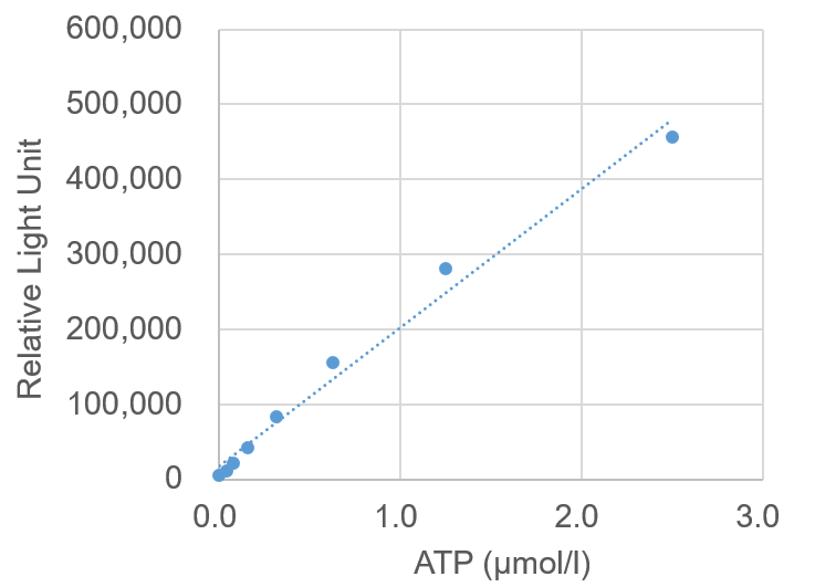
Preparation of Calibration Curve
ATP levels in a sample can be measured by a calibration curve established with ATP Standards included in this kit. If the ATP levels are greater than or equal to 2.5 µmol/l, the sample must be diluted before measurement.
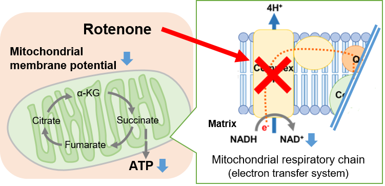
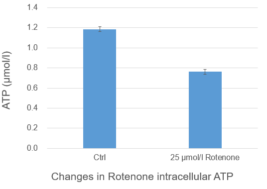
Change in intracellular metabolism of rotenone-treated cells
Rotenone, which is known to inhibit the mitochondrial electron transport chain, was added to Jurkat cells, followed by measurement of intracellular ATP using the ATP Assay Kit-Luminescence. As a result, ATP production in the mitochondrial respiratory chain (the electron transport chain) was inhibited, and ATP concentrations were lower than those in the control cells.
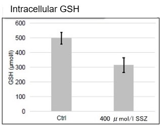
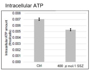
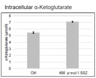
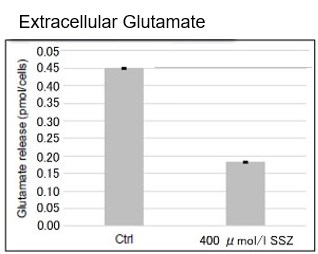
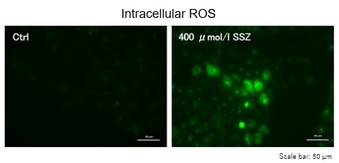
Sulfasalazine Alters Intracellular Metabolites and Increases ROS in A549 Cells
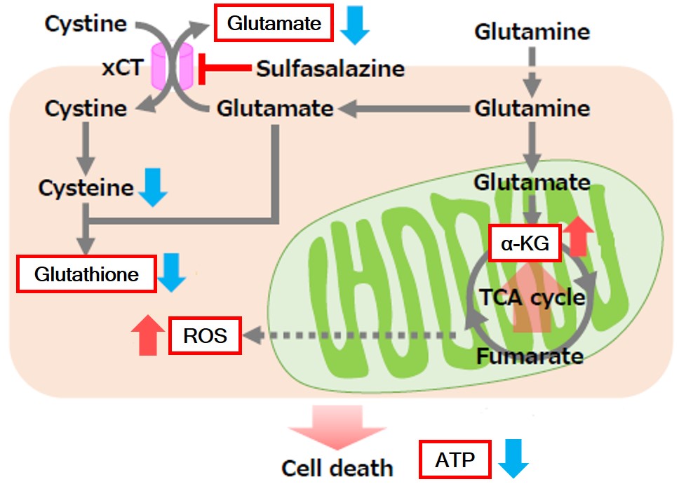 |
After addition of sulfasalazine (SSZ), a known inhibitor of cystine/glutamate transporter (xCT), to A549 cells, we observed changes in intracellular glutathione (GSH), ATP, α-ketoglutarate (α-KG), ROS and glutamate release. The results indicated that the addition of SSZ
|
     |
|
Experimental Example: Change in Metabolism in Liver Tissue of MASH-Induced Mouse
It is known that metabolic dysfunction-associated steatohepatitis (MASH) results in decreased ATP, α-ketoglutarate (α-KG), and NAD levels in affected tissues. ATP, α-KG, and NAD levels were measured in the liver tissue of type 1 diabetic model mice (STAM model) that were treated with a high-fat diet (MASH induction) since 4 weeks old. Measurement at 10 weeks old confirmed that ATP, α-KG, and NAD levels indeed decreased in the tissue samples after MASH induction.
Note: for more details about the experimental procedure, please refer to Q&A "Are there any examples of experiments using tissue samples?"
・Cellular ATP :ATP Assay Kit-Luminescence (Code:A550).
・Cellular α-KG:α-Ketoglutarate Assay Kit-Fluorometric (Code:K261).
・Cellular NAD :NAD/NADH Assay Kit-WST (Code:N509).
References:
| ATP | Francesco Bellanti, et al., "Synergistic interaction of fatty acids and oxysterols impairs mitochondrial function and limits liver adaptation during nafld progression", Redox Biology, 2018, 15, 86-96. |
|---|---|
| α-KG | Jianjian Zhao, et al., "The mechanism and role of intracellular α-ketoglutarate reduction in hepatic stellate cell activation", Bioscience Reports, 2020, 40, (3). |
| Ali Canbay, et al., "L‑Ornithine L‑Aspartate (LOLA) as a Novel Approach for Therapy of Non‑alcoholic Fatty Liver Disease", Drugs, 2019, 79, 39-44. | |
| NAD | Jinhan He, et al., "Activation of the Aryl Hydrocarbon Receptor Sensitizes Mice to Nonalcoholic Steatohepatitis by Deactivating Mitochondrial Sirtuin Deacetylase Sirt3", Mol. and Cell. Biol., 2013, 33, (10), 2047-55. |
References
| No. | Sample | Instrument | Reference (Link) |
|---|---|---|---|
| 1) | Cell (DLD-1) |
Plate Reader |
G. Enkhbat, A. Nakanishi, Y. Miki, "The BRCA2 missense mutation K2497R suppressed self-degradation and increased ATP production and cell proliferation", 2021, doi:10.1016/j.bbrc.2021.12.073. |
Q & A
-
Q
How many samples can I measure?
-
A
The number of samples that can be recorded when the standard curve and sample are measured in triplicates. (50 tests/kit: 8 samples, 200 tests/kit: 48 samples; two times measurement for preparation of standard curves) Please refer to the instruction manual for a 96-well plate layout.
-
Q
How about the stability of luminescence signals?
-
A
A luminescence signal remains stable for 3 hours. However, luminescence is affected by temperature and light. If you cannot measure luminescence immediately, please keep the plate in the dark and maintain a constant temperature of around 25℃.
-
Q
May I use color plates other than white plates for measurement?
-
A
Yes, you may. However, luminescence intensity decreases if you use black or transparent plates. Strong luminescent signals are detected in blank wells particularly in transparent plates. Therefore, it is strongly recommended to use white plates.
We have tested with the following white microplates.
Company Product name Bottom Cat No. SUMITOMO BAKELITE White plate for luminescence measurement with lid White
MS-8096WZ Thermo Scientific 96 Well White/Clear Bottom Plate, TC Surface Clear 165306 SPL Life Sciences White plate Clear 33696 Greiner Greiner Bio-One CELLSTAR 96-well,
Cell Culture-Treated, Flat-Bottom MicroplateClear 655098
-
Q
Can I store my samples after an experimental treatment?
-
A
No. You cannot store them because ATP is unstable. Please add a Working solution to each well immediately after an experimental treatment and measure them within 3 hours.
-
Q
Can I store the Working solution?
-
A
Yes. After the preparation, the Working solution is stable at - 20 ℃ for approximately 30 days. Avoid freeze-thaw cycles to prevent degradation of the solution; aliquot it into micro tubes and store at -20ºC.
-
Q
Are there any examples of experiments using tissue samples?
-
A
Yes. We have examples using mouse liver tissue.
General protocol is as below.
Extraction of ATP from liver samples via alkaline extraction method
1)Add cold 0.5 mol/l KOH solution to mouse liver (500 µl /100 mg).
*Make sure blood is properly removed from the tissue sample, using hemoperfusion or other methods.
*Any residual blood may affect the measurement.
2) Homogenize the tissue with Dounce tissue grinder.
3) Collect the sample and transfer it to a sample tube.
Wash the tissue grinder with 500 µl of cold 0.5 mol/l KOH solution and transfer the solution into the sample tube. (Total volume: 1 ml)
4) Add 1 ml of cold ultrapure water to the sample tube, mix well, and put it on ice for 5 minutes. (Total volume: 2 ml)
*If the solution is highly viscous, it may be difficult to separate after centrifugation in the next step.
In this case, attach a needle (about 25 g) to a syringe and use the syringe to pipette the sample solution about 20-30 times until the
sample solution can be smoothly pipetted within the syringe.
5) Centrifuge at 12,000 x g , 4 °C for 5 minutes and collect each 900 µl of the supernatant in two new tubes.
Preparation of sample for ATP measurement
6) Add 200 µl of 1 mol/l KH2PO4 solution to 900 µl of the sample prepared in step 5. Mix well and put it on ice for 5 minutes.
7) Centrifuge at 12,000 x g for 5 minutes at 4°C and collect 1 ml of the supernatant in a tube as a sample for measurement.
*The extracted tissue samples cannot be stored. Please measure the sample within a day of the extraction.
*For dilution of the “standard” and “sample”, use a mixture of 0.5 mol/l KOH solution and 1 mol/l KH2PO4 solution at a ratio of 9:5 as the dilution solution.
ATP Level Changes in the Liver Tissue of NASH-Induced Mice
-
Q
What is the wavelength of the luminescence measurement?
-
A
This kit uses firefly luciferin. Therefore, the luminescence measurement wavelength is 556 nm.
Handling and storage condition
| -20℃, Protect from moisture |


