

|
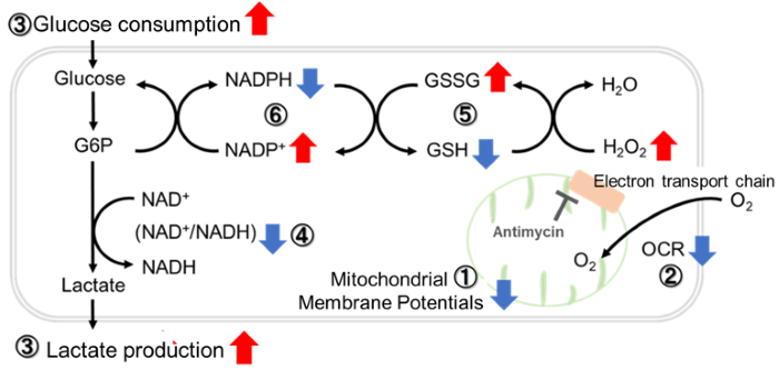
Mitochondrial metabolism involves the biochemical processes within mitochondria that convert nutrients into energy and building blocks necessary for cell function, primarily through the citric acid cycle and oxidative phosphorylation. This energy production produces adenosine triphosphate (ATP), the cell's primary energy currency. Oxidative stress occurs when there's an imbalance between the production of reactive oxygen species (ROS) in the mitochondria and the cell's ability to detoxify these harmful byproducts or repair the resulting damage. Over time, excessive oxidative stress can lead to cellular damage that contributes to aging and several diseases, including neurodegenerative disorders and cancer. |
|
| Related Techniques |
|
|
|
|
|
|
|
|
| Related Applications |
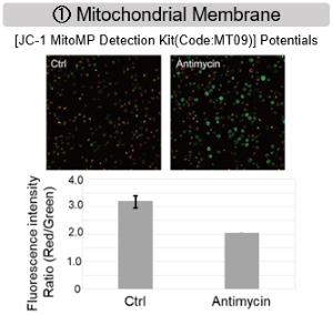
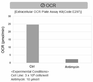
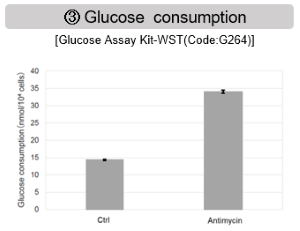
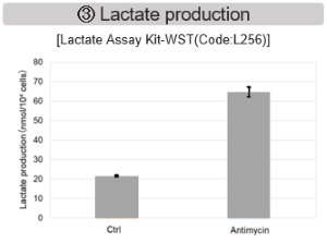
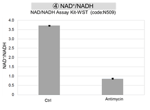
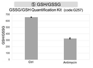
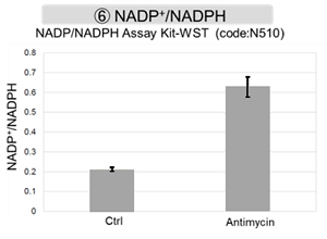
Inhibition of Mitochondrial Electron Transport Chain
|
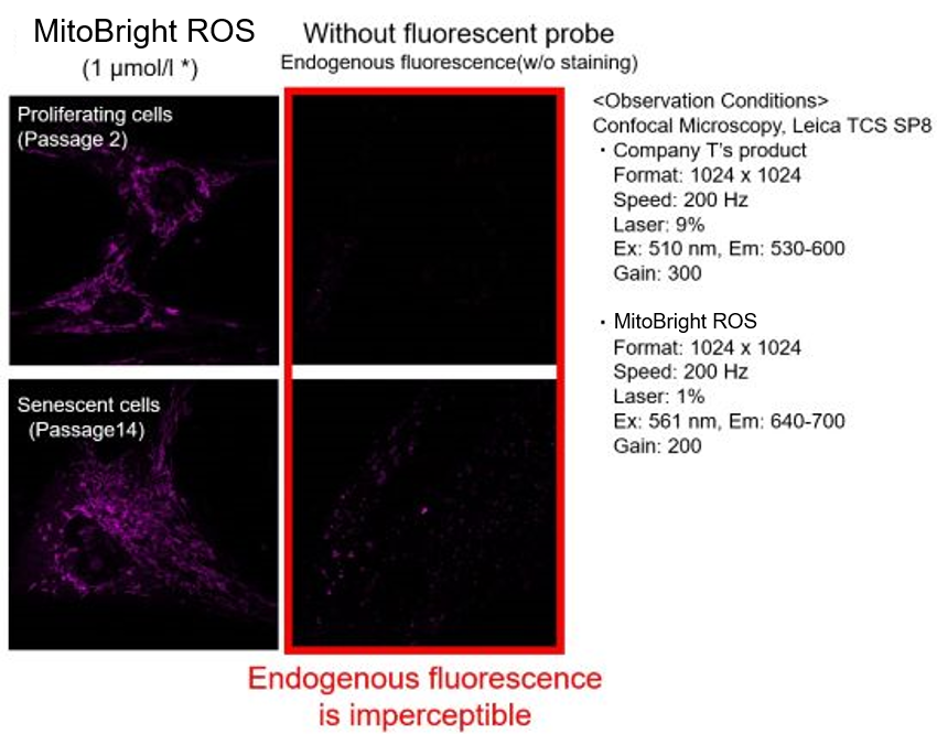
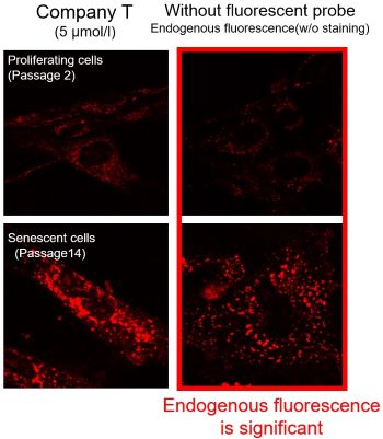
Mitochondrial Superoxide Detection in Senescent CellsBackground fluorescence caused by lipofuscin can be minimized by using a better fluorescent probe, as tested in TIG-1 cells. Lipofuscin accumulates in senescent cells, causing increased background fluorescence during observation. To minimize the effects of endogenous fluorescence from lipofuscin and other substances, a better fluorescent probe was tested in TIG-1 cells. Company T's product exhibited endogenous fluorescence, while MitoBright ROS Deep Red showed less background fluorescence. Researchers should compare sensitivity, wavelength, and channels and select the appropriate fluorescent probe to minimize endogenous fluorescence for accurate cellular senescence research. Products in Use |


Product Classification
Product Classification
-
Cell Proliferation / Cell Cytotoxicity Assay
Cell Proliferation / Cell Cytotoxicity Assay Kits /Related Reagents
-
Cell Staining
Cell Double Staning Kit /Live Cell Staining /Dead Cell Staining /Nuclear Staining /Mitochondria Staning /Tissue Staining /Nucleolus Staining /Lipid Droplet Staining /Cell Membrane Staining /Lysosome Staining
-
Intracellular Fluorescent Probes
Reagents for Intracellular Calucuum Ion /Reagents for Intracellular Ion /Related Reagents
-
Labeling Chemistry
Protein Labeling Kits /Protein Labeling Reagents /HPLC Derivertization Reagents /Biotion Labeling Reagents /Related Reagents /Exsosome Labeling
-
Oxidative Stress
Stress Maker Detection /NO Detection /NO Donor /NO Inhibitor /ACE Inhibition Assay /Reagents・Kits for Sulfur Biology /Antioxidant Assay Kit /Donors for Sulfur Biology
-
-Bacstain- Series
Bacterial Proliferation Assay Kit /Bacteria Staining /Bacterial Fluorescent Staining
-
Molecular Biology
Transfection Reagents /Nuclear Staining /Agarose /Related Reagents /Buffer for Molecular Biology
-
Detergents
Detergents /Sets
-
Cross-Linking Reagents
Hetero-bifunctional Reagents /Homo-bifunctional Reagents /Others
-
Redox Dyes
Reductive Chromogenic Dyes /Electron Mediators /Oxidative Chromogenic Dyes /Trinder Reagents
-
Ion Analysis
Ionophores /Anion Eliminator /Solvent for Ion Electrode of Liquid Film Type
-
Organic Scintillator
-
Buffers
Buffers
-
Metal Chelates
EDTA /Other Chelator /Reagents for Chelator Titration
-
Chromogen/Metal Indicator
Chromogen/Metal Indicator
-
Water Analysis
/Fluorine /Iron / /Water Hardness /Residual Chlorine /ABS /Cyan / /Chromium /Copper
-
Extraction Reagent
AA Chelator /Related Reagents
-
High Purity Solvent
Spectrozole /Luminazole / /Acnazole /Dehydration Solvent for Synthesis
-
Biochemicals
Biochemicals
-
Functional Organic Material
Alkanethiol Derivative /Phosphonic Acid Derivatives
