|
Through single-cell RNA sequencing (scRNA-seq), they identified an indispensable transcription factor, CBFb that ensures AM self-renewal. The study concludes that AMs in aged hosts exhibit signs of senescence, possibly due to reduced activity of the CBFb transcription factor. |
|||
|
Single cell RNA sequencing unravels mechanisms underlying senescence-like phenotypes of alveolar macrophages |
|||
|
Point of Interest |
|||
| Related Techniques | |||
| First choice for cellular senescence assay | Cellular Senescence Detection Kit – SPiDER-ßGal | ||
| Cellular senescence assay with a plate reader | Cellular Senescence Plate Assay Kit – SPiDER-ßGal | ||
| Cell cycle assay | Cell Cycle Assay Solution Blue / Deep Red | ||
| Dead cell staining for flow cytometry | Dead Cell Makeup Blue / Deep Red - Higher Retention than PI | ||
| Lysosomal pH detection | Lysosomal Acidic pH Detection Kit-Green/Red, Green/Deep Red | ||
| Mitochondrial function/glycolysis detection | Glycolysis/JC-1 MitoMP Assay Kit | ||
| Oxygen consumption rate assay | Extracellular OCR Plate Assay Kit | ||
| Related Applications | |||
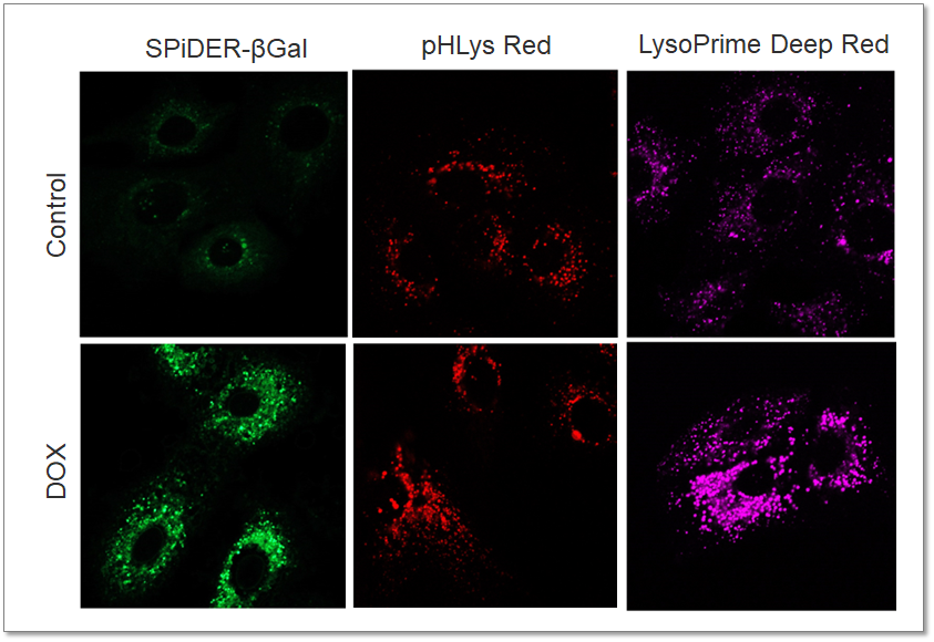
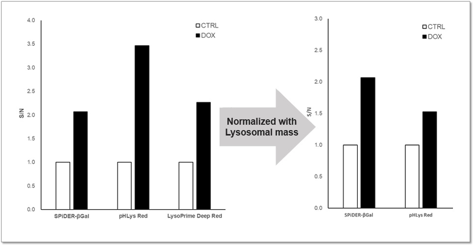
| Analysis of Lysosomal Mass and pH Exchange in Senescence-induced Cells | |||
|
|
Purpose: To investigate changes in lysosomal mass and pH in A549 cells induced to senescence by treatment with Doxorubicin (DOX). Methods: Senescence-associated acidic β-galactosidase (SA-βGal) activity was detected using Cellular Senescence Detection Kit - SPiDER-βGal. Lysosomal mass was detected using LysoPrime Deep Red, and pH was detected using pHLys Red. Fluorescence imaging was used to observe changes in lysosomal mass and pH in senescent cells compared to non-senescent cells. The normalized fluorescence intensity of lysosomal mass and pH was also measured by a plate reader. Results: Our findings indicate that senescence induced by DOX resulted in an increase in lysosomal mass and acidification of pH compared to non-senescent cells. The obtained results are consistent with previous reports* that demonstrated enhanced lysosomal activity in senescent cells induced by the CDK4/6 inhibitor, palbociclib. The fluorescence imaging and plate reader data both support these findings. * Miguel Rovira, et. al., Aging Cell (2022)
<Experimental Conditions for Microscopy> <Experimental Conditions for Plate Reader>
<Products in Use> |
||