

|
This article focusing on the mechanisms of programmed necrosis in infected macrophages. The research demonstrates that the excess tumor necrosis factor triggers programmed necrosis of infected macrophages is not mitochondrion-intrinsic but results from an inter-organellar circuit initiating and culminating in the mitochondrion. The circuit begins and ends with the transit of two inorganic signals - ROS from mitochondrion to lysosome and Ca2+ from endoplasmic reticulum to mitochondrion - and requires cathepsin D translocation from lysosome to cytosol. |
|
TNF Induces Pathogenic Programmed Macrophage Necrosis in Tuberculosis through a Mitochondrial-Lysosomal-Endoplasmic Reticulum Circuit Click here for the original article: Francisco J. Roca, et. al., Cell (2019) Point of Interest
|
| Related Techniques |
|
|
|
|
|
|
|
| Related Applications |
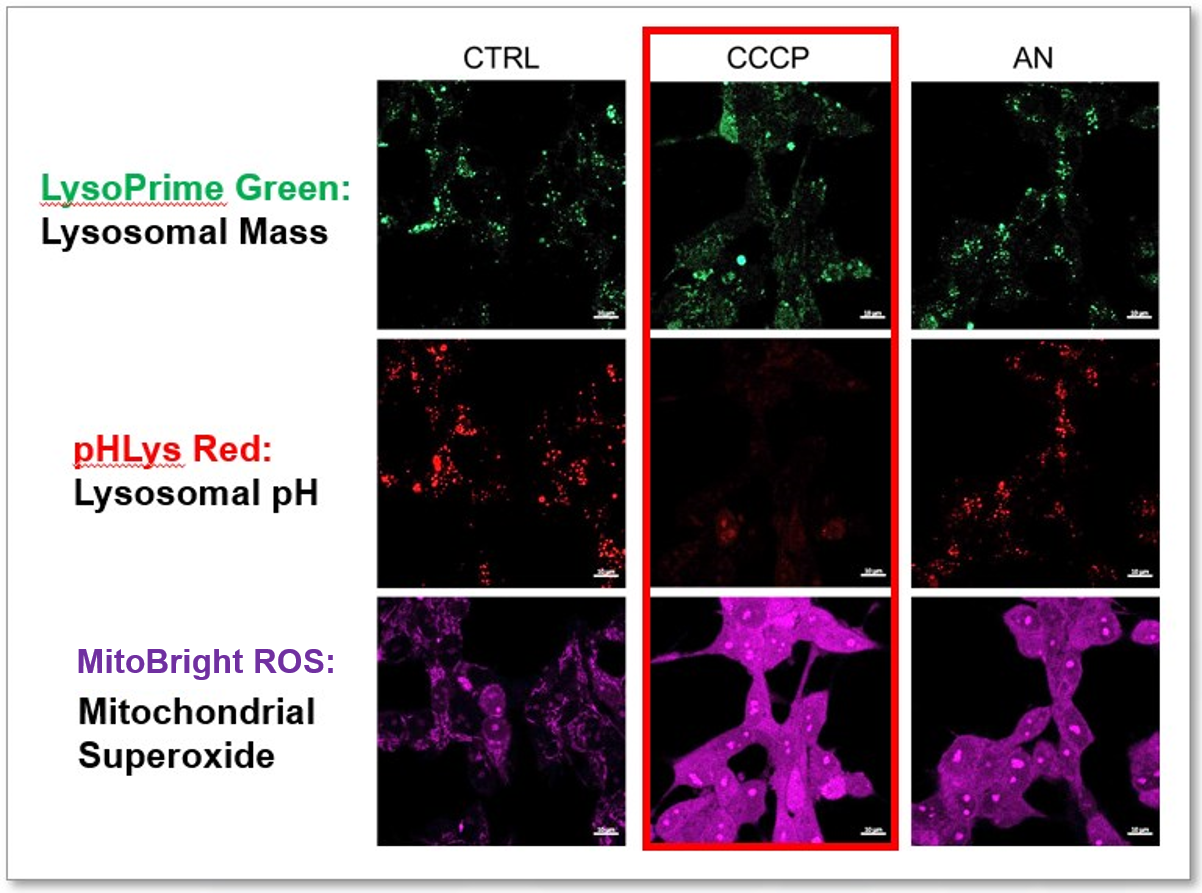
1. Simultaneously detection of Lysosomal and Mitochondrial Dysfunction
|
|
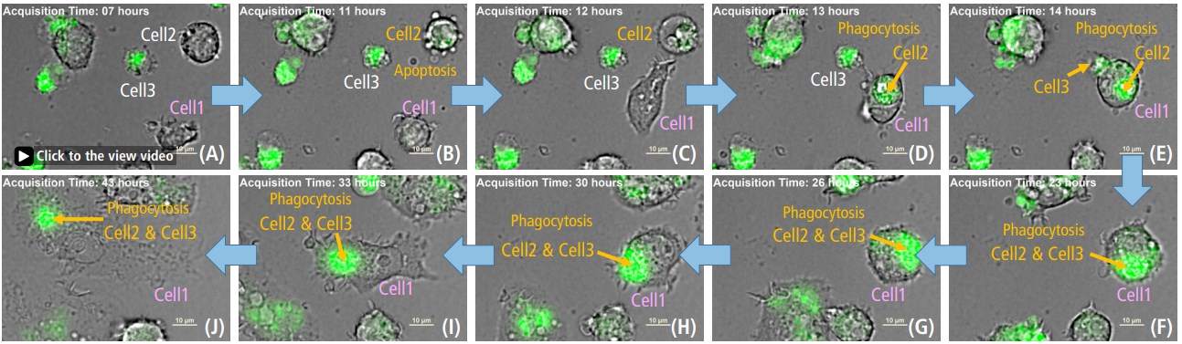
2. Monitoring ROS in Macrophage Phagocytosis Dead cells (2&3) phagocytosed by Cell1 resulted in increased ROS(green).
ROS detection reagent allowed for reliable analysis of the role of ROS in phagocytosis. Its high intracellular residence and low background noise made it possible to perform long-term analysis of ROS production in the cell. This information can provide important insights into the mechanisms of phagocytosis and contribute to the development of treatments for diseases associated with macrophage dysfunction. > for detail experimental notes are available at Nikon web site. Products in Use Related Product |


Product Classification
Product Classification
-
Cell Proliferation / Cell Cytotoxicity Assay
Cell Proliferation / Cell Cytotoxicity Assay Kits /Related Reagents
-
Cell Staining
Cell Double Staning Kit /Live Cell Staining /Dead Cell Staining /Nuclear Staining /Mitochondria Staning /Tissue Staining /Nucleolus Staining /Lipid Droplet Staining /Cell Membrane Staining /Lysosome Staining
-
Intracellular Fluorescent Probes
Reagents for Intracellular Calucuum Ion /Reagents for Intracellular Ion /Related Reagents
-
Labeling Chemistry
Protein Labeling Kits /Protein Labeling Reagents /HPLC Derivertization Reagents /Biotion Labeling Reagents /Related Reagents /Exsosome Labeling
-
Oxidative Stress
Stress Maker Detection /NO Detection /NO Donor /NO Inhibitor /ACE Inhibition Assay /Reagents・Kits for Sulfur Biology /Antioxidant Assay Kit /Donors for Sulfur Biology
-
-Bacstain- Series
Bacterial Proliferation Assay Kit /Bacteria Staining /Bacterial Fluorescent Staining
-
Molecular Biology
Transfection Reagents /Nuclear Staining /Agarose /Related Reagents /Buffer for Molecular Biology
-
Detergents
Detergents /Sets
-
Cross-Linking Reagents
Hetero-bifunctional Reagents /Homo-bifunctional Reagents /Others
-
Redox Dyes
Reductive Chromogenic Dyes /Electron Mediators /Oxidative Chromogenic Dyes /Trinder Reagents
-
Ion Analysis
Ionophores /Anion Eliminator /Solvent for Ion Electrode of Liquid Film Type
-
Organic Scintillator
-
Buffers
Buffers
-
Metal Chelates
EDTA /Other Chelator /Reagents for Chelator Titration
-
Chromogen/Metal Indicator
Chromogen/Metal Indicator
-
Water Analysis
/Fluorine /Iron / /Water Hardness /Residual Chlorine /ABS /Cyan / /Chromium /Copper
-
Extraction Reagent
AA Chelator /Related Reagents
-
High Purity Solvent
Spectrozole /Luminazole / /Acnazole /Dehydration Solvent for Synthesis
-
Biochemicals
Biochemicals
-
Functional Organic Material
Alkanethiol Derivative /Phosphonic Acid Derivatives
