|
Ferroptosis and senescence are two distinct cellular processes that significantly impact disease initiation and progression. Ferroptosis is an iron-dependent form of cell death characterized by lipid peroxidation that plays a critical role in neurodegenerative diseases by promoting neuronal death and in cancer where it can either inhibit or promote tumor growth depending on the context. Senescence involves a state of permanent cell cycle arrest that acts as a tumor suppressor by preventing the proliferation of damaged cells, but also contributes to aging and tissue dysfunction. Both processes are integral to the pathophysiology of several diseases and provide potential targets for therapeutic intervention.
|
-
Aging promotes metabolic dysfunction-associated steatotic liver disease by inducing ferroptotic stress
Click here for the original article: Kuo Du, et. al., Nature Aging, 2024.
Point of Interest
- A gene cluster characteristic of aging hepatocytes is enriched in diseased livers and includes genes that induce ferroptosis.
- Aged mice show increased hepatocyte ferroptosis and liver degeneration under conditions that induce metabolic stress.
- Inhibition of ferroptosis in aged mice reverses age-related liver damage.
-
Microglial ferroptotic stress causes non-cell autonomous neuronal death
Click here for the original article: Jeffrey R. Liddell et. al., Mol Neurodegener, 2024.
Point of Interest
- Ferroptotic stress in microglia triggers inflammatory cascades, leading to neuronal death through neurotoxic astrocytes.
- Human amyotrophic lateral sclerosis (ALS) spinal cord tissue shows ferroptosis signatures, that are also observed in the ALS mouse model, where ferroptosis inhibition is neuroprotective.
- Ferroptosis in microglia can cause non-cell autonomous neuronal death, suggesting a novel pathophysiological role in neurodegenerative diseases.
-
Fatty acid binding protein 5 suppression attenuates obesity-induced hepatocellular carcinoma by promoting ferroptosis and intratumoral immune rewiring
Click here for the original article: Jonathan Sun et. al., Nature Metabolism, 2024.
Point of Interest
- BRCA1 deficiency in cancers confers resistance to erastin-induced ferroptosis but increases susceptibility to GPX4 inhibitor-induced
- Fatty acid binding protein 5 (FABP5) drives obesity-induced hepatocellular carcinoma (HCC), and its inhibition induces lipid peroxidation and ferroptosis in cancer cells.
- FABP5 inhibition reduces HCC burden in mice by promoting a pro-inflammatory tumor microenvironment.
- FABP5 inhibition activates CD8+ T cells and increases expression of CD80 and CD86, T cell activation molecules in tumor-associated macrophages.
|
|
Related Techniques
|
- Intracellular / mitochondrial ferrous ion (Fe2+) detection
- FerroOrange(intracellular), Mito-FerroGreen(mitochondrial)
|
- Cellular senescence detection
- SPiDER-βGal for live-cell imaging or flow cytometry / microplate reader / tissue samples
SPiDER-βGal Blue for fixed cell and for multiple staining with immunostaining and other methods
|
|
|
- Lipid Peroxidation Assay
- Lipid Peroxidation Probe -BDP 581/591 C11-
|
- Total ROS detection
- Highly sensitive DCFH-DA or Photo-oxidation Resistant DCFH-DA
|
- Mitochondrial superoxide detection
- MitoBright ROS Deep Red - Mitochondrial Superoxide Detection
|
- Mitochondrial membrane potential detection
- JC-1 MitoMP Detection Kit, MT-1 MitoMP Detection Kit
|
- Glutathione Quantification
- GSSG/GSH Quantification Kit
|
- Cystine Uptake detection
- Cystine Uptake Assay Kit
|
- MDA detection
- MDA Assay Kit
|
- Mitophagy or autophagy detection
- Mitophagy Detection Kit, Autophagic Flux Assay Kit
|
- Lysosomal function
- Lysosomal Acidic pH Detection Kit -Green/Red and Green/Deep Red
|
- Glycolysis/Oxidative phosphorylation Assay
-
- Glycolysis/OXPHOS Assay Kit
-
|
- Apoptosis detection in multiple samples
-
- Annexin V Apoptosis Plate Assay Kit
-
|
|
Related Applications
|
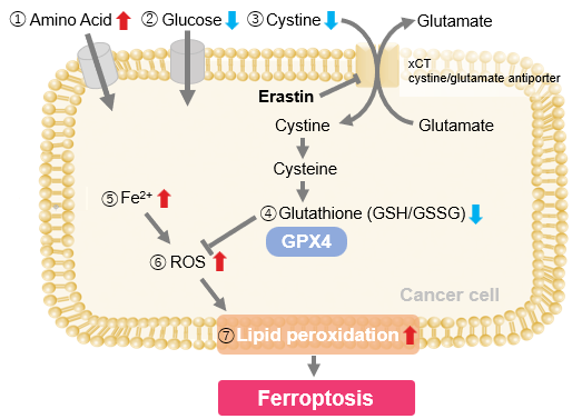
Erastin-Induced Ferroptosis: Evaluating Intracellular Uptake and Redox Balance
We investigated the transition of cellular metabolisms in A549 cells treated with erastin, a known ferroptosis inducer. Our results revealed the following.

- The inhibition of cystine uptake by erastin led to a depletion of cysteine, which in turn increased the compensatory uptake of other amino acids.
- Glucose uptake, which typically promotes ferroptosis*, was found to decrease upon erastin treatment, suggesting a potential cellular self-defense mechanism.
- The depletion of cysteine resulted in a decrease in glutathione levels and an increase in Fe2+, ROS, and lipid peroxides, all of which are recognized markers of ferroptosis.
Cell Line: A549
Incubation Conditions: 100 μmol/l Erastin/MEM, 37℃, 3h
*Reference: Xinxin Song, et al., Cell Reports, (2021)
Products in Use
|


