


Lysosomal Lipid Radical Probe -Lyso-NBD-Pen-
Lysosomal Lipid Radical Detection
- Enables detection of lipid radicals in lysosomes of live cells
- Compatible with fluorescence microscopy and flow cytometry for detection and quantification
- Detects changes in lipid localization after reaction through binding to lipid radicals
-
Product codeL271 Lysosomal Lipid Radical Probe -Lyso-NBD-Pen-
| Unit size | Price | Item Code |
|---|---|---|
| 2 nmol | $380 | L271-10 |
*2 nmol can be used for 10 assays in 35 mm dishes (final concentration: 0.1 µmol/l).

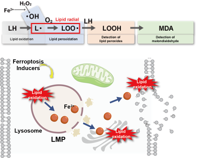
The Importance of Lipid Radicals in Ferroptosis
Ferroptosis is a form of cell death triggered by iron-dependent lipid peroxidation and has been increasingly implicated in various diseases, including cancer, neurodegenerative diseases, and age-related disorders. Lipid radicals are key intermediates involved in the propagation of lipid peroxidation chain reactions and are attracting attention as indicators of early changes and progression of lipid oxidation during ferroptosis. In recent years, ferroptosis has also been reported to be initiated by lysosomal lipid peroxidation, highlighting the importance of detecting lipid radicals in lysosomes.¹⁾
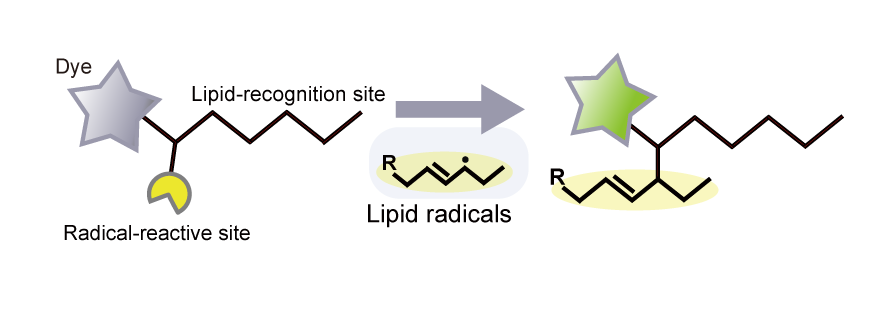
Lyso-NBD-Pen localizes to lysosomes and enables specific detection of lipid radicals generated within lysosomes. Simply add the reagent to cultured cells to detect lysosomal lipid radicals using fluorescence microscopy or flow cytometry.
1) S.Saimoto, et al., Nat. Commun., 2025, 16, 3554.
This product was developed under the guidance of Professor Kenichi Yamada of Kyushu University.
| Product Name | Target | Detection Properties |
|---|---|---|
| FerroOrange | Intracellular Fe2+ | Microscopy, Plate reader Ex: 543 nm / Em: 580 nm |
| Mito-FerroGreen | Mitochondrial Fe2+ | Microscopy Ex: 505 nm / Em: 535 nm |
| Lyso-FerroRed | Lysosomal Fe2+ | Microscopy, FCM, Plate reader Ex: 551 nm / Em: 571 nm |
| Iron Assay Kit -Colorimetric- | Fe2+ and Fe3+ | Plate reader Colorimetric, λ: 593 nm |
| Liperfluo | Lipid Peroxide | Microscopy, FCM Ex: 488 nm / Em: 500-550 nm |
| Lipid Peroxidation Probe -BDP 581/591 C11- |
Lipid Peroxidation Process |
Microscopy, FCM, Plate reader |
| MDA Assay Kit | Malondialdehyde | Plate reader Fluorescence, Ex: 540 nm / Em: 590 nm Colorimetric, λ: 532 nm |
| Cystine Uptake Assay Kit | Cystine uptake | Plate reader Ex: 485 nm / Em: 535 nm |
| GSSG/GSH Quantification Kit II | GSSG and GSH | Plate reader Colorimetric, λ: 405 nm |

Manual
Technical info
Please select the appropriate reagent based on your experimental method and detection instrument used for lipid peroxidation analysis.
| Liperfluo | [This Product] Lysosomal Lipid Radical Probe -Lyso-NBD-Pen- | Lipid Radical Probe -NBD-Pen- | Lipid Peroxidation Probe -BDP 581/591 C11- | |
| Target | Lipid peroxides | Lipid radicals | Lipid radicals | Lipid radicals + hydroxyl radicals |
|
Localization |
Intracellular |
Lysosomal |
Intracellular |
Intracellular |
|
Compatible Instruments |
Fluorescence microscopy, |
Fluorescence microscopy, |
Fluorescence microscopy, Flow cytometry |
Fluorescence microscopy, |
|
Detection Conditions |
Fluorescence |
Fluorescence Ex: 488 nm, Em: 490–600 nm |
Fluorescence Ex: 488 nm, Em: 490–600 nm |
Fluorescence |
| Sensitivity | ☆ | ☆☆ | ☆☆☆ | |
|
Interaction with Target |
Reacts only |
Reacts and binds |
Reacts only (does not bind) |
|
| Features | Specifically detects lipid peroxides. |
Binds to lipid radicals, enabling detection of changes in lipid localization after the reaction. |
Ratiometric detection enables quantification using a plate reader. | |
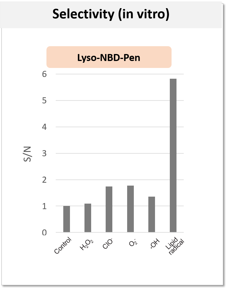
High Selectivity for Lipid Radicals
The reactive oxygen species listed below were mixed with this reagent in 100 mmol/l phosphate buffer (pH 7.4), and the fluorescence intensity was measured after a 30-minute reaction.
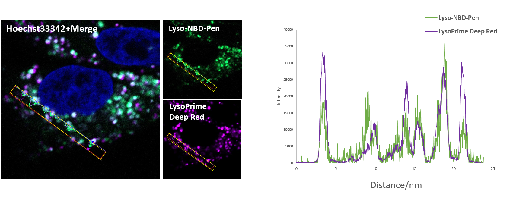
Reliable Lysosomal Localization
To confirm the lysosomal localization of this probe reagent, co-staining was performed with the lysosome staining reagent LysoPrime Deep Red (Product Code: L264). The fluorescence signals of both reagents were colocalized, confirming that Lyso-NBD-Pen selectively stains lysosomes.
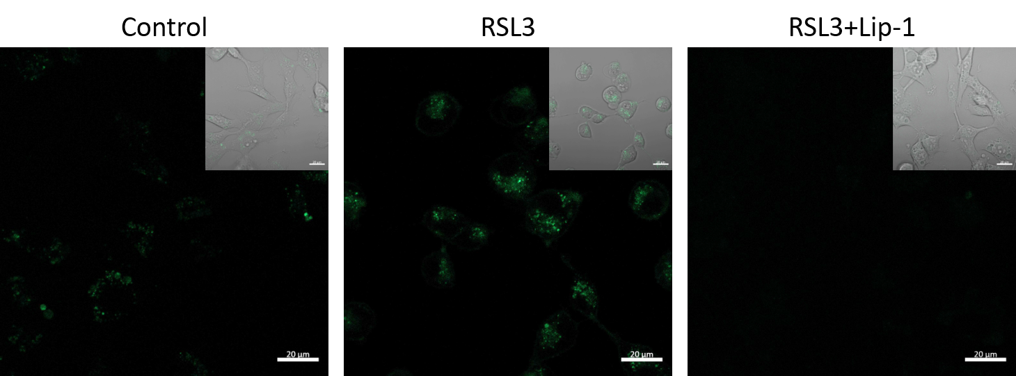
Detection of Lysosomal Lipid Radicals
Treatment with the ferroptosis inducer RSL3 resulted in an increase in lipid radicals within lysosomes. Furthermore, co-treatment with the lipid radical scavenger Liproxstatin-1 (Lip-1) resulted in a decrease in the signal, indicating that this reagent selectively detects lipid radicals.
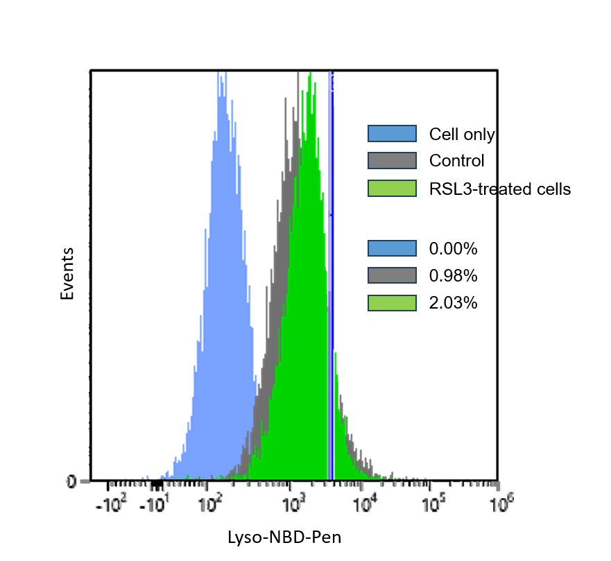
Quantification of Lysosomal Lipid Radicals
HT1080 cells were treated with the ferroptosis inducer RSL3, and the increase in lysosomal lipid radicals was quantified by flow cytometry.
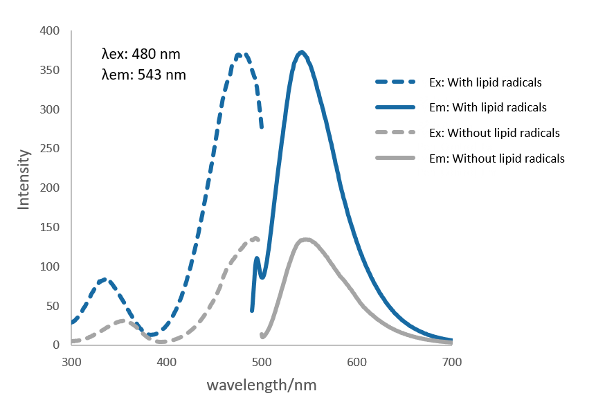
Fluorescence Properties
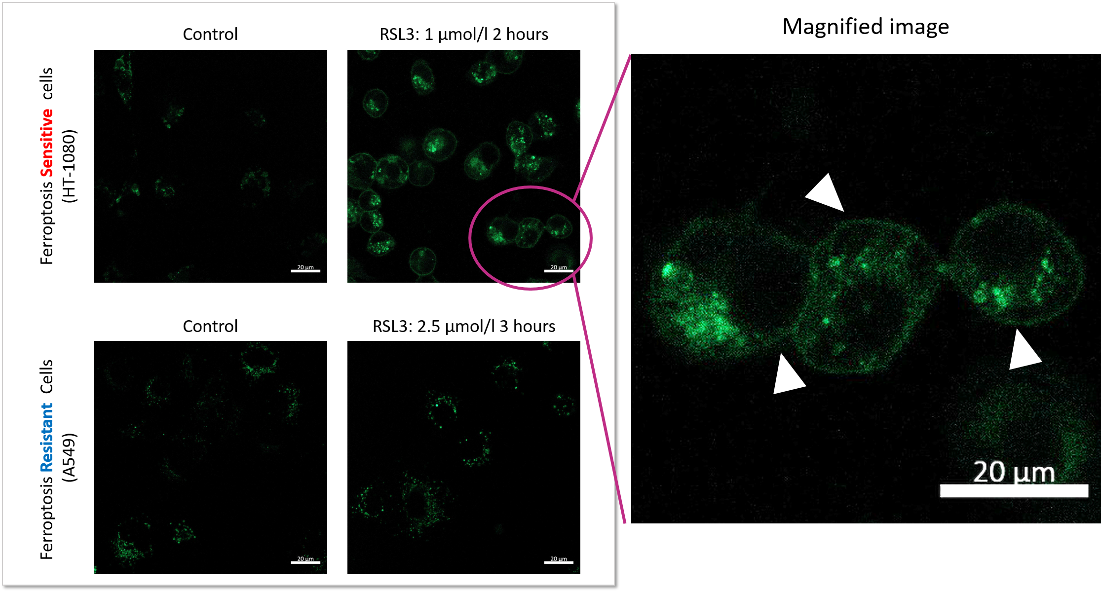
Experimental Example: Comparison of Lysosomal Lipid Radical Detection in Ferroptosis-Sensitive and -Resistant Cells
Changes in lysosomal lipid radicals were detected in HT-1080 cells, which are highly sensitive to ferroptosis, and A549 cells, which are resistant to ferroptosis, following treatment with RSL3, a ferroptosis inducer.
In A549 cells, a ferroptosis-resistant cell line, no significant difference in fluorescence intensity was observed compared with the control, even when the RSL3 treatment concentration and duration of RSL3 exposure were increased (concentration: up to 2.5 μmol/l; duration: up to 3 hours). In contrast, in HT-1080 cells, a ferroptosis-sensitive cell line, treatment with 1 μmol/L RSL3 for 2 hours increased fluorescence derived from lysosomal lipid radicals and caused changes in their localization.
These results demonstrate that this product can detect differences in lysosomal lipid radical generation and localization changes depending on the cell type.
Q & A
-
Q
How many samples can be measured with this product?
-
A
The number of samples that can be measured with 2 nmol of this product is as follows:
- 96-well plate: 2 plates
- ibidi 8-well plate: 100 wells
- 35 mm dish: 10 dishes
- 6-well plate: 10 wells
-
Q
Is there an experimental example that can be used as a positive control?
-
A
Please refer to the instruction manual for the lysosomal lipid radical detection experiment using HT-1080 cells treated with RSL3 to induce ferroptosis.
If endogenous lipid radicals are detected, they can be scavenged by co-treatment with Liproxstatin-1.
-
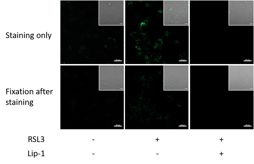
Q
Can Lyso-NBD-Pen be used for observation after cell fixation following staining?
-
A
No. This product is suitable only for live-cell imaging, as the fluorescence signal decreases after fixation.
-
Q
What precautions should be taken when observing samples with a fluorescence microscope?
-
A
NBD, the fluorescent scaffold of this probe, undergoes photobleaching when continuously exposed to excitation light.
When acquiring imaging data, adjust the focus under bright-field observation before acquiring fluorescence images.
-
Q
How long can samples be observed after staining?
-
A
We have confirmed that samples can be observed for up to 24 hours after staining.
Handling and storage condition
| Appearance: | Orange to red solid |
|---|---|
| Solubility in Dimethyl sulfoxide: | To pass test |
| Dye content: | To pass test |
| Store at room temperature. |


