 Hidden sections will not be printed.
General Information
Ferroptosis is a form of cell death caused by the abnormal accumulation of lipid peroxides in an iron‑dependent manner. During the progression of lipid peroxidation, various reaction intermediates are generated as a result of lipid oxidation. A recent report indicates that ferroptosis is initiated by lipid peroxidation of the lysosomal membrane, highlighting the importance of detecting lipid radicals within lysosomes1).
Lyso‑NBD‑Pen is a fluorescent probe that localizes to lysosomes and selectively reacts with lipid radicals. When added to cultured cells, Lyso‑NBD‑Pen permeates the cell membrane, accumulates in lysosomes, and emits a fluorescent signal upon reaction with lipid radicals, enabling the detection of lysosomal lipid radicals in living cells.
Content
| Lysosomal Lipid Radical Probe -Lyso-NBD-Pen- | 2 nmol |
Storage Condition
Store in a cool and dark place.
Required Equipment and Materials
- Fluorescence microscope, or flow cytometer
- Incubator (37℃)
- Micropipettes (100–1000 µl, 20–200 µl, 1–10 µl)
- Microtubes
- Medium or Hanks' Balanced Salt Solution (HBSS)
Precaution
- Centrifuge the tube briefly before opening the cap because the contents may adhere to the tube wall or the inside of the cap.
- Please refer to Table 1 for suitable fluorescence wavelengths for each application.
Table 1. Recommended filter settings for Lyso-NBD-Pen
| Applications | Fluorescence microscope | Flow cytometer |
| Measurement wavelength |
・Confocal microscope Ex/Em: 488/490–600 nm ・Fluorescence microscope GFP filter |
FITC filter |
Preparation of Solutions
Preparation of 100 µmol/l Lyso-NBD-Pen DMSO stock solution
Add 20 μl of DMSO to a tube containing 2 nmol of Lyso-NBD-Pen and dissolve by pipetting to prepare a 100 µmol/l Lyso-NBD-Pen DMSO stock solution.
- The 100 µmol/l Lyso-NBD-Pen DMSO stock solution is stable at −20℃ for 1 month.
Preparation of 100 nmol/l Lyso-NBD-Pen working solution
Dilute the 100 µmol/l Lyso-NBD-Pen DMSO stock solution 1:1000 in medium to prepare 100 nmol/l Lyso-NBD-Pen working solution.
- The working solution cannot be stored and must be prepared each day freshly.
- Refer to Table 2 for the amount of working solution required by vessel type.
Table 2. Required amount of the working solution by vessel type
| Vessel | 35-mm dish | ibidi 8-well plate | 96-well black plate (clear bottom) |
| Appropriate amount | 2 ml | 200 μl/well | 100 μl/well |
General Protocol
- Seed cells in a vessel. Culture the cells at 37°C overnight in a 5% CO2 incubator.
- Discard the supernatant and wash the cells once with medium.
- Discard the supernatant, add an appropriate volume of 100 nmol/l Lyso-NBD-Pen working solution to the vessel, and incubate at 37°C for 30 min in a 5% CO2 incubator.
- Discard the supernatant and wash the cells twice with medium.
- Add medium containing ferroptosis inducers or inhibitors, and incubate the cells at 37°C in a 5% CO2 incubator for an appropriate time.
- Discard the supernatant and wash the cells twice with HBSS.
- Observe the cells under a fluorescence microscope or measure fluorescence signals using a flow cytometer.
Usage Example 1
Detection of lysosomal lipid radicals in HT-1080 cells treated with RSL3 using a confocal laser microscope
- HT-1080 cells (3×10⁴ cells/well) in MEM (supplemented with 10% fetal bovine serum and 1% penicillin–streptomycin) were seeded in an ibidi 8-well plate and incubated at 37°C overnight in a 5% CO2 incubator.
- After the supernatant was removed, the cells were washed once with medium. Then, 100 nmol/l Lyso-NBD-Pen working solution (200 µl/well) was added to the cells, and they were incubated at 37°C for 30 min in a 5% CO2 incubator.
- The supernatant was removed, and the cells were washed twice with medium.
- RSL3 (1 µmol/l) dissolved in medium was added to the cells, and they were incubated at 37°C for 2 h in a 5% CO2 incubator.
- The supernatant was removed, and the cells were washed twice with HBSS. The wells were refilled with HBSS.
- The cells were observed under a confocal laser microscope.
 |
Detection: Confocal laser microscope |
Figure 1. Fluorescence images of HT-1080 cells obtained with a confocal laser microscope
Usage Example 2
Detection of lysosomal lipid radicals in HT-1080 cells treated with RSL3 using a flow cytometer
- HT-1080 cells (3×10⁵ cells/well) in MEM (supplemented with 10% fetal bovine serum and 1% penicillin–streptomycin) were seeded in a 6-well plate and incubated at 37°C overnight in a 5% CO2 incubator.
- After the culture medium was removed, the cells were washed once with medium. Then, 100 nmol/l Lyso-NBD-Pen working solution (2 ml/well) was added to the cells, and they were incubated at 37°C for 30 min in a 5% CO2 incubator.
- The supernatant was removed, and the cells were washed twice with medium.
- RSL3 (1 µmol/l) dissolved in medium was added to the cells, and they were incubated at 37°C for 2 h in a 5% CO2 incubator.
- After removing the supernatant, the cells were washed once with HBSS and detached from the plate using 0.25% trypsin–EDTA.
- The detached cells were collected into a 1.5‑ml tube containing serum-supplemented medium and centrifuged at 300 × g for 5 min.
- The supernatant was discarded, 1 ml of PBS was added to resuspend the cells, and the cells were centrifuged at 300 × g for 5 min.
- The supernatant was discarded, and the cells were resuspended in 1 ml of PBS.
- The samples were passed through a cell strainer for flow cytometry and analyzed using a flow cytometer.
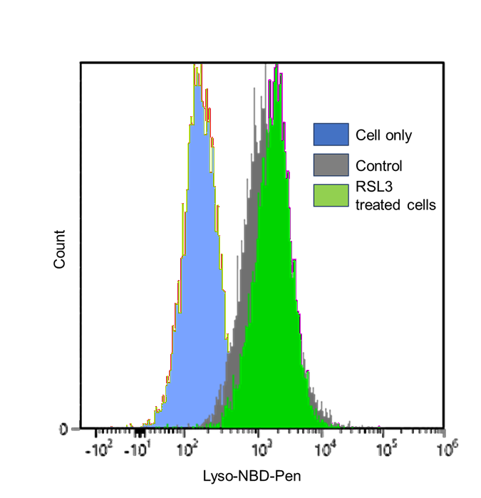 |
Detection: Flow cytometer |
Figure 2. Fluorescence signals from HT-1080 cells, detected using a flow cytometer
Reference
1) K. Yamada et al., Nature Communications, 2025, 16, 2554.
Frequently Asked Questions / Reference


L271: Lysosomal Lipid Radical Probe -Lyso-NBD-Pen-
Revised May., 15, 2026