

|
Recent cancer research has focused on intratumoral senescent cancer cells and it is becoming clear that these cells contribute to immune evasion of cancer cells and create a cancer-promoting environment. Here are some of the papers that have identified senescent cells or senescent cells with specific phenotypes in cancer treatment and have shown that removing these cells is important for cancer treatment. Senescent cells are damaged or stressed cells that permanently stop dividing but do not undergo cell death. In the context of cancer, senescent cells can accumulate in tumors, particularly after treatments such as chemotherapy, contributing to an immunosuppressive environment. This environment can promote tumor growth, immune evasion and resistance to therapy. However, the selective targeting and elimination of senescent cells, known as senolytic therapy, is emerging as a promising strategy to improve cancer treatment by reducing tumor progression and improving patient outcomes. |
|||
|
The efficacy of chemotherapy is limited by intratumoral senescent cells expressing PD-L2 |
Cellular senescence in malignant cells promotes tumor progression in mouse and patient Glioblastoma |
Senescence drives immunotherapy resistance by inducing an immunosuppressive tumor microenvironment Click here for the original article: Damien Maggiorani, et. al., Nature Communications, 2024. |
|
|
Point of Interest - PD-L2 is not necessary for senescence but crucial for immune evasion by senescent cells, promoting tumor persistence after chemotherapy. - Antibody-mediated PD-L2 blockade synergizes with chemotherapy, suggesting a potential therapeutic strategy targeting senescent cancer cells. |
Point of Interest - Removal of p16Ink4a-expressing senescent cells from GBM tumors improves survival in mice and alters the tumour environment. - Senolytic drugs that target senescent cells may be a promising adjuvant therapy for GBM patients, improving survival. |
Point of Interest - Elimination of senescent cells restores CD8 T cell proliferation and reduces immunotherapy resistance. - Anti-senescent cell drugs prior to immune checkpoint inhibitors may enhance the effectiveness of cancer immunotherapy. |
|
| Related Techniques | |||
| Cellular senescence detection | SPiDER-βGal for live-cell imaging or flow cytometry / microplate reader / tissue samples NEW SPiDER-βGal Blue for fixed cell and for multiple staining with immunostaining and other methods |
||
| Glycolysis/Oxidative phosphorylation Assay | Glycolysis/OXPHOS Assay Kit | ||
| Endocytosis Detection detection | ECGreen-Endocytosis Detection | ||
| Lysosomal function | Lysosomal Acidic pH Detection Kit-Green/Red and Green/Deep Red | ||
| First-time autophagy research | Autophagic Flux Assay Kit | ||
| Total ROS detection | Highly sensitive DCFH-DA or Photo-oxidation Resistant DCFH-DA | ||
| Apoptosis detection in multiple samples | Annexin V Apoptosis Plate Assay Kit | ||
| Cell proliferation/ cytotoxicity assay | Cell Counting Kit-8 and Cytotoxicity LDH Assay Kit-WST | ||
| Glutathione Quantification | GSSG/GSH Quantification Kit | ||
| Related Applications | |||
Co-staining with Lipid droplet and SA-β-Gal in fixed cells |
|||
|
|
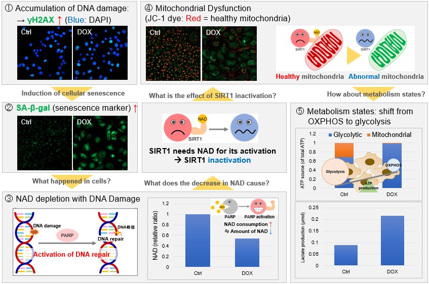
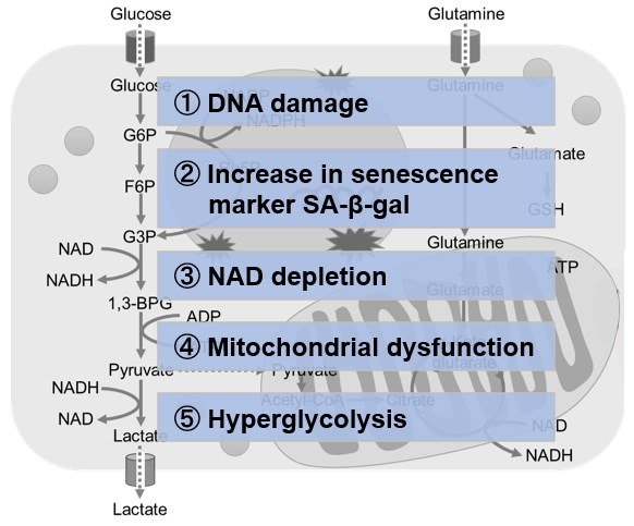
NAD(+) levels decline during the aging process, causing defects in nuclear and mitochondrial functions and resulting in many age-associated pathologies*. Here, we try to redemonstrate this phenomenon in the doxorubicin (DOX)-induced cellular senescence model with a comprehensive analysis of our products. *S. Imai, et al., Trends Cell Biol, 2014, 24, 464-471
|
||
 |
|||
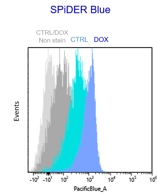
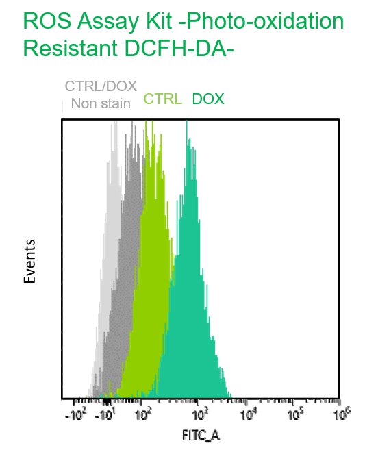
Multiple staining with oxidative stress-related markers using Doxorubicin-induced senescent cells(flow cytometry) |
|||
|
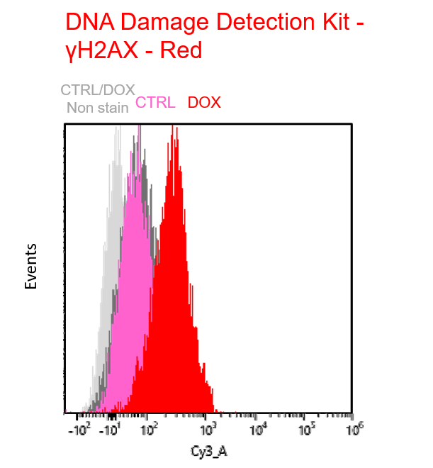
Using A549 cells induced to senescence by doxorubicin (DOX) and normal cells (CTRL), changes in oxidative stress-related markers in senescent cells were analyzed by flow cytometry with multiple staining. SA-βGal as a senescence marker was detected by Cellular Senescence Detection Kit - SPiDER Blue, total ROS as an oxidative stress marker was detected by ROS Assay Kit - Photo-oxidation Resistant DCFH-DA-, and γH2AX as a DNA damage marker was detected by DNA Damage Detection Kit - γH2AX-Red. As a result, total ROS and γH2AX were increased in SA-βGal-positive senescent cells, and the increase in oxidative stress-related markers associated with cellular senescence could be detected by multiple staining. <Experimental Procedure> |
|||


Product Classification
Product Classification
-
Cell Proliferation / Cell Cytotoxicity Assay
Cell Proliferation / Cell Cytotoxicity Assay Kits /Related Reagents
-
Cell Staining
Cell Double Staning Kit /Live Cell Staining /Dead Cell Staining /Nuclear Staining /Mitochondria Staning /Tissue Staining /Nucleolus Staining /Lipid Droplet Staining /Cell Membrane Staining /Lysosome Staining
-
Intracellular Fluorescent Probes
Reagents for Intracellular Calucuum Ion /Reagents for Intracellular Ion /Related Reagents
-
Labeling Chemistry
Protein Labeling Kits /Protein Labeling Reagents /HPLC Derivertization Reagents /Biotion Labeling Reagents /Related Reagents /Exsosome Labeling
-
Oxidative Stress
Stress Maker Detection /NO Detection /NO Donor /NO Inhibitor /ACE Inhibition Assay /Reagents・Kits for Sulfur Biology /Antioxidant Assay Kit /Donors for Sulfur Biology
-
-Bacstain- Series
Bacterial Proliferation Assay Kit /Bacteria Staining /Bacterial Fluorescent Staining
-
Molecular Biology
Transfection Reagents /Nuclear Staining /Agarose /Related Reagents /Buffer for Molecular Biology
-
Detergents
Detergents /Sets
-
Cross-Linking Reagents
Hetero-bifunctional Reagents /Homo-bifunctional Reagents /Others
-
Redox Dyes
Reductive Chromogenic Dyes /Electron Mediators /Oxidative Chromogenic Dyes /Trinder Reagents
-
Ion Analysis
Ionophores /Anion Eliminator /Solvent for Ion Electrode of Liquid Film Type
-
Organic Scintillator
-
Buffers
Buffers
-
Metal Chelates
EDTA /Other Chelator /Reagents for Chelator Titration
-
Chromogen/Metal Indicator
Chromogen/Metal Indicator
-
Water Analysis
/Fluorine /Iron / /Water Hardness /Residual Chlorine /ABS /Cyan / /Chromium /Copper
-
Extraction Reagent
AA Chelator /Related Reagents
-
High Purity Solvent
Spectrozole /Luminazole / /Acnazole /Dehydration Solvent for Synthesis
-
Biochemicals
Biochemicals
-
Functional Organic Material
Alkanethiol Derivative /Phosphonic Acid Derivatives
