ROS Research

Compatible with Immunostaining

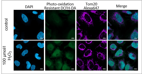
Co-staining with anti-protein immunostain

After adding hydrogen peroxide, HeLa cells were stained with Photo-oxidation resistant DCFH-DA and mitochondrial marker Tom 20. As a result, the intracellular ROS level and mitochondrial morphology were clearly observed at the same time, which is very difficult for the existing ROS probe.

2. ROS Assay Kit - Highly Sensitive DCFH-DA

Super High-Sensitive ROS Detection

Comparison of Sensitivity: Microscope           
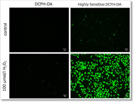
                                        

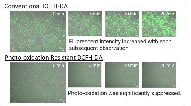
Hydrogen peroxide (H2O2)-treated HeLa cells (1×10⁴ cells/ml) were stained with DCFH-DA or the ROS Assay Kit-Highly Sensitive DCFH-DA, and the detectability of intracellular ROS was compared between two detection kits.  Results indicated that the ROS Assay Kit-Highly Sensitive DCFH-DA was better at high-sensitivity detection of intracellular ROS than regular DCFH-DA in high-sensitivity detection of intracellular ROS was better than DCFH-DA.

Ex: 450-490 nm, Em: 500-550 nm (GFP filter), Scale bar: 50 μm


 Comparison of Sensitivity: Microscope

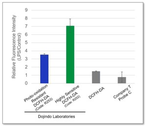
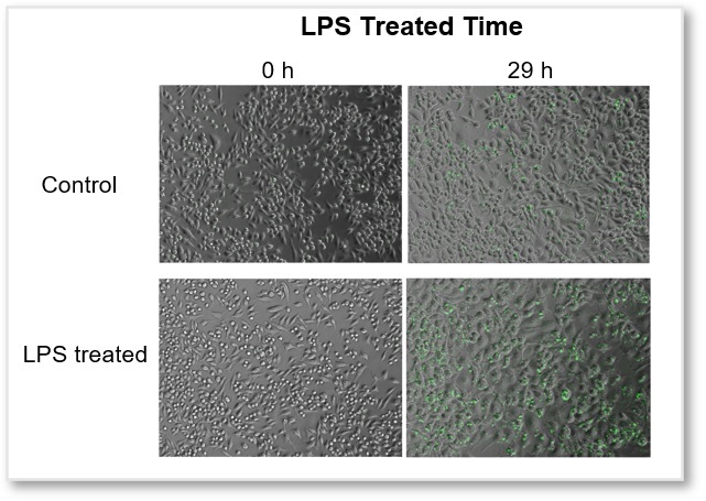
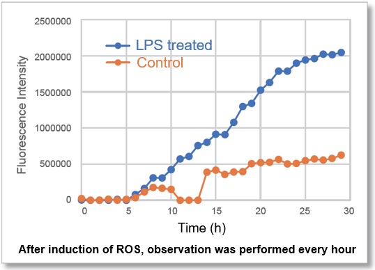
In Lipopolysaccharide (LPS)-treated RAW 264.7 cells, after being stained with regular DCFH-DA, Highly Sensitive DCFH-DA, or Photo-oxidation Resistant DCFH-DA, the intracellular ROS levels were compared. The results showed that the Dojindo Laboratories’ probes could detect intracellular ROS with higher sensitivity.

Ex: 490 – 520 nm, Em:  510 – 540 nm


Application Data

Simultaneously evaluation of mitochondrial superoxide and intracellular total ROS

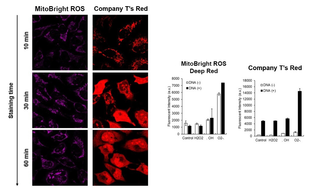
HeLa cells were washed with HBSS and co-stained with MitoBright ROS Deep Red - Mitochondrial Superoxide Detection and ROS Assay Kit -Highly Sensitive DCFH-DA- (Total ROS detection), and separately treated with mitochondrial superoxide inducer Antimycin or hydrogen peroxide. 

As a result, intracellular total ROS and mitochondrial superoxide were observed separately.


General Protocol

Treated with H2O2


Treated with Antimycin

<Imaging Conditions>(Confocal microscopy)
Intracellular ROS: Ex = 488, Em = 490-520 nm
MitoBright ROS: Ex = 633 nm, Em = 640-700 nm
Scale bar: 10 μm


Disruption of cystine balance changes nutrients uptake and redox level

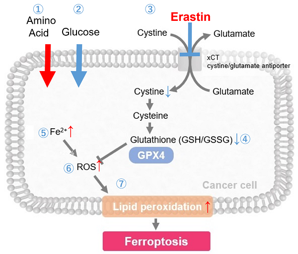
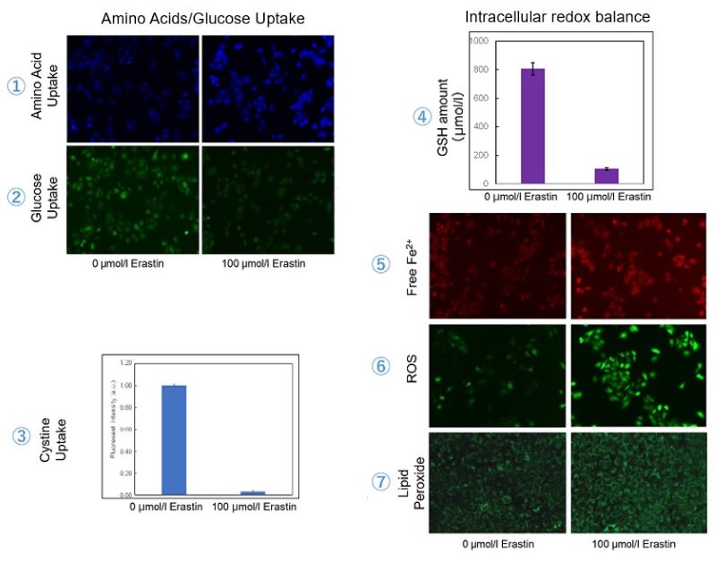
Cystine/glutamate antiporter (xCT) is frequently overexpressed in human cancers and promotes cystine uptake and glutathione biosynthesis*. This results in protection from oxidative stress and ferroptosis, a type of programmed cell death dependent on iron and characterized by the accumulation of lipid peroxides. We, therefore, treated A549 cells with erastin to inhibit xCT before evaluating intracellular uptake and redox balance

Pranavi Koppula, et. al., Cancer Commun., 38:12 (2018)


As a result, we were able to get three conclusions:
・To compensate for the loss of cystine, the uptake of amino acids increased.
・The decrease in glucose uptake suggests that metabolic reprogramming may have taken place.
・The decrease in glutathione would have increased Fe2+, ROS, and Lipid peroxides – all hallmarks of Ferroptosis.


Related Products

Product

Unit

Code

Cystine Uptake Assay Kit

20 tests/100 tests

UP05-10/UP05-12

Amino Acid Uptake Assay Kit

20 tests/100 tests

UP04-10/UP04-12

Glucose Uptake Assay Kit-Blue

100 tests

UP01-10

Glucose Uptake Assay Kit-Green

20 tests/100 tests

UP02-10/UP06-10

Glucose Uptake Assay Kit-Red

100 tests

UP03-10

GSSG/GSH Quantification Kit

200 tests

G257-10

FerroOrange

1 tube/3 tubes

F374-10/F374-12

ROS Assay Kit -Highly Sensitive DCFH-DA-

100 tests

R252-10

MitoBright ROS Deep Red - Mitochondrial Superoxide Detection 100 nmol x1/100 nmol x 3 MT16-10/MT14-12

Liperfluo

50 µg x 5

L248-10

Product Classification

Product Classification