


Glucose Uptake Assay Kit-Blue
- Cellular function
- Cellular senescence
- Cellular metabolism
- Fluorescent dye
- Fluorescence microscope
- FCM
Glucose Uptake Capacity Assay
- Highly sensitive and simple measurement of glucose uptake capacity
- Applicable for microscopy & FCM
- Reduces dye leakage after staining
-
Product codeUP01 Glucose Uptake Assay Kit-Blue
| Unit size | Price | Item Code |
|---|---|---|
| 1 set | $486.00 | UP01-10 |
General number of usable assays for 1 set
35 mm dish x 12 or 96-well microplate x 1


| 1 set | Glucose Uptake Probe-Blue WI Solution (50×) |
×1 5 ml ×1 |
|---|
Description
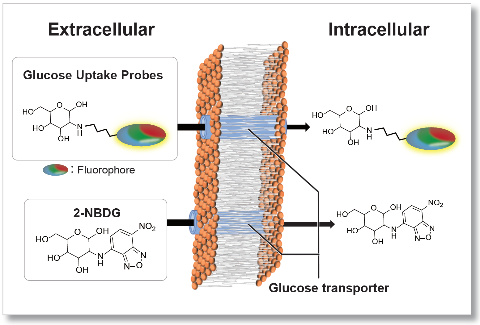
Nutrient metabolism is necessary for energy production in cells and regulates various cellular functions, including gene expression. Glucose is one of the key substrates for the generation of ATP and sustaining cellular homeostasis. Thus, glucose metabolism has been the subject of intense investigations. In cancer research, tumor cells enhance glucose uptake and consumption for their growth and proliferation. Therefore, elevated glucose uptake is a marker of tumors, and glucose transporters are important targets in cancer treatment.
One standard method for evaluating the glucose uptake ability of cells uses radioisotope-labeled glucose. Although this method has been used for many years, it requires special handling facilities and disposal of radioactive materials. The enzyme cycling method using 2-deoxy-D-glucose, which enables colorimetric and fluorometric plate assays, cannot be applied to cell imaging and flow cytometry. Recently, 2-NBDG, a fluorescently labeled glucose analog, has been used widely to detect cellular glucose uptake by fluorescence imaging and flow cytometry. However, the sensitivity of this method is poor because of the low fluorescence intensity of 2-NBDG.
To resolve these limitations, novel fluorescent probes, Glucose Uptake Probes, were developed. These probes emit strong fluorescence, allowing highly sensitive detection of cellular glucose uptake by fluorescence imaging, flow cytometry, or microplate assay. The WI Solution in these kits enhances the cellular retention of the probe to give more reliable data.

Glucose Uptake Assay Products
| Products | Plate Reader | Microscope / FCM |
|---|---|---|
| Glucose Uptake Assay Kit-Blue | ✗ | ✓ |
| Glucose Uptake Assay Kit-Green | ✓ | ✓ |
| Glucose Uptake Plate Assay Kit (Green) | ✓ Best* | ✗ |
| Glucose Uptake Assay Kit-Red | ✓ | ✓ |
*No washing step, minimizing cell stress and detachment while improving reproducibility.


Manual
Technical info
Four Advantages Compare to Existing Method
Glucose Uptake Probe-Blue allowing highly sensitive detection of cellular glucose uptake by fluorescence imaging or flow cytometry. The WI Solution in this kit can enhance cellular retention to provide more reliable experimental data. Also, compare with the existing method (2-NBDG), the measurement time can be significantly reduced.
①Multi-color Imaging
Glucose Uptake Probe can easily co-stain with Green or red or any other long-wavelength fluorescent reagent. The following image is a co-staining result with Glucose Uptake Probe-Blue and Lipid Droplets (Red: Lipi-Red, Code LD03) in adipocytes differentiated 3T3-L1 cells.

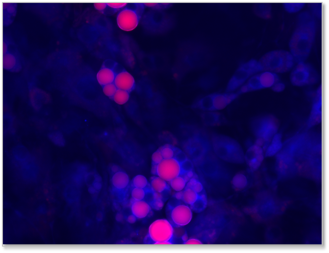
<Experimental Conditions>
Cell line: 3T3-L1
Imaging Conditions
Glucose Uptake Probe-Blue: Ex=340-380 nm, Em=435-485 nm
Lipi-Red: Ex=533-557 nm, Em=570-640 nm
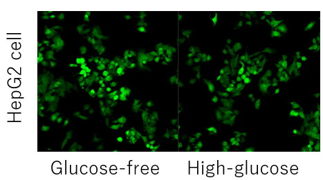
②Microscopy & FCM Compatibility
Glucose Uptake Probe-Blue is compatible with microscope and Flow Cytometer. The result below is using Glucose Uptake Probe-Blue to evaluate A549 cells' glucose uptake capability with No Glucose and High Glucose medium.


Cell Line: A549
Imaging Conditions:
Glucose Uptake Assay Kit-Blue: Ex=340-380 nm, Em=435-485 nm

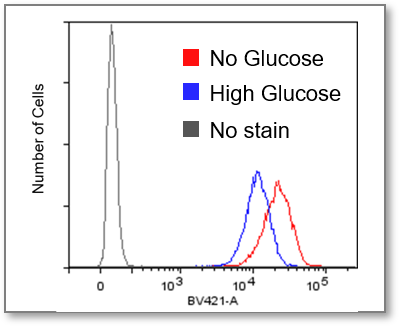
Cell Line: A549
Experimental Conditions:
Glucose Uptake Assay Kit-Blue: Ex=405 nm, Em=425-475 nm
③Quick Process
Significantly reduced measurement time despite using the same process as 2-NBDG.
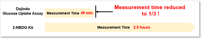
The operation is very simple, requiring only three steps: pretreatment, staining (incorporation), and washing.
.png)
④Reduces Dye Leakage After Staining
The WI Solution in this kit can enhance cellular retention to provide more reliable experimental data.
For more details, please refer to the page of (Glucose Uptake Assay Kit-Green, Code: UP02).
Comparison with Existing Method
The comparison of the Glucose Uptake Probe Series and the existing method(2-NBDG) is as below.
| product name | Fluorescence microscope | Plate reader detection | FCM detection | Retention ability | Fluorescence characteristics |
|---|---|---|---|---|---|
| Glucose Uptake Assay Kit-Blue | ✓ | - | ✓ | 1 hour * | λex:386 nm λem:474 nm |
| Glucose Uptake Assay Kit-Green | ✓ | ✓ | ✓ | 1 hour * | λex:507 nm λem:518 nm |
| Glucose Uptake Assay Kit-Red | ✓ | ✓ | ✓ | 1 hour * | λex:560 nm λem:572 nm |
| 2-NBDG | ✓ | - | ✓ | 30 minutes or less * | λex:465 nm λem:540 nm |
*Result of A549 cells, the retention time for other cell lines may be different.
Experimental Example: Inhibition of Glucose Uptake by Cytochalasin B
Using Glucose Uptake Probe Series to detect the inhibition of glucose uptake by the glucose transporter inhibitor Cytochalasin B in HepG2 cells with high sensitivity.
Imaging with microscopy

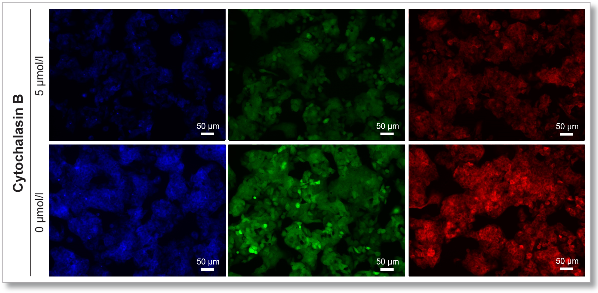
(Scale bar: 50 μm)
<Experimental Conditions>
Cell Line: HepG2
Miedum: MEM (5.5 mmol/l Glucose)
Incubation Conditions: 5 µmol/l Cytochalasin B / MEM (5.5 mmol/l Glucose, 10% FBS), 37℃, 24 hr
Reagent Conditions: Glucose Uptake Probe (500x dilution)/DMEM (0 mol/l Glucose), 37℃, 15 min
Device: Fluorescent Microscopy
Glucose Uptake Assay Kit-Blue: Ex=340-380 nm, Em: 435-485 nm
Glucose Uptake Assay Kit-Green: Ex=450-490 nm, Em: 500-550 nm
Glucose Uptake Assay Kit-Red: Ex=533-557 nm, Em: 570-640 nm
Experimental Example: Enhanced Glucose Uptake by Insulin
Using Glucose Uptake Probe to evaluate insulin-induced glucose uptake enhancing in adipocytes with high sensitivity.
Imaging with microscopy


(Scale bar: 50 µm)
<Experimental Conditions>
Cell: mouse adipocyte
Miedum: DMEM (5.5 mmol/l Glucose, 10% FBS)
Inducer Conditions:0 or 1 µmol/l Insulin / DMEM (0 mmol/l Glucose , serum-free), 37℃, 15 min
Reagent Conditions:Glucose Uptake Probe (500x dilution) /DMEM (0 mmol/l Glucose, serum-free), 37℃, 15 min
Device: Fluorescent Microscopy
Glucose Uptake Assay Kit-Blue: Ex=340-380 nm, Em: 435-485 nm
Glucose Uptake Assay Kit-Green: Ex=450-490 nm, Em: 500-550 nm
Glucose Uptake Assay Kit-Red: Ex=533-557 nm, Em: 570-640 nm
Please refer to the link below for details regarding Insulin treatment.

Application Data: Starvation induced changes in autophagy and glucose uptake
HeLa cells were stained with autophagosome staining reagent DAPRed and autolysosome staining reagent DALGreen and then starved in an amino acid-free medium for 3 hours to induce autophagy. The fluorescence intensity of DAPGreen and DALGreen are increased which indicated autophagy, and the increase in glucose uptake capacity of the cells was also observed using Glucose Uptake Probe-Blue.
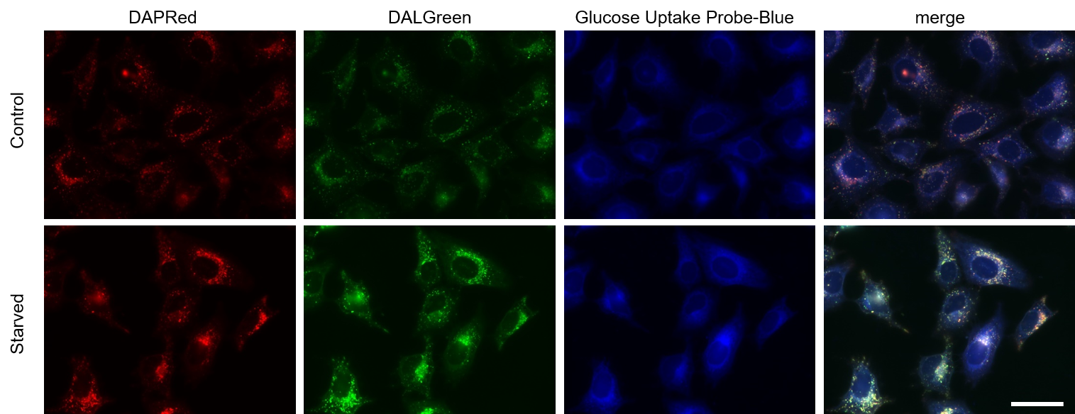
Fluorescent Microscope
Blue: Ex = 340-380 nm, Em = 435-485 nm
Green: Ex = 450-490 nm, Em = 500-550 nm
Red: Ex = 533-557 nm, Em = 570-640 nm
Scale bar: 50 μm
Difference from Glucose Assay Kit
The difference between Glucose Uptake Assay Kit and Glucose Assay Kit-WST(Code: G264)
1. Glucose Assay Kit-WST can quantify the glucose concentration in cell supernatants.
Glucose Uptake Assay Kit does not apply for the quantification of glucose.
2. Glucose Uptake Assay Kit can evaluate cellular glucose uptake capability in a short time.
Glucose Assay Kit-WST can not measure the glucose level changes in a short period of time.
For more details, please refer to the page of Glucose Uptake Assay Kit-Green (Code: UP02).
Q & A
-
Q
Does Glucose Uptake Probe-Blue have selectivity for the type of glucose transporter?
-
A
No. Glucose Uptake Probe-Blue is thought to be taken up by cells via multiple glucose transporters. However, there are no detailed data on the selectivity and affinity of this probe for glucose transporters.
-
Q
Which cell lines have been analysed with this kit?
-
A
We have tested the kit in the following cell lines.
Cell lines Human alveolar basal epithelial adenocarcinoma cells A549 Human hepatoma-derived cells HepG2 Progenitor adipocytes Preadipocytes (3T3-L1) Adipocytes Adipocytes (3T3-L1) Lewis lung cancer-derived cells 3LL T cells CD4+ T cell
-
Q
Which inhibitor or experiments altering cellular uptake capacity have been tested with this kit?
-
A
Cell line Dilution factor of probe stock solution staining time Examples Human alveolar basal epithelial adenocarcinoma cells A549 x 500 15分 Phloretin, WZB117 Human hepatoma-derived cells HepG2 x 500 15分 Cytochalasin B Preadipocytes preadipocyte(3T3-L1) x 500 15分 Differentiation to adipocytes Adipocytes adipocyte(3T3-L1) x 500 15分 Insulin Lewis lung cancer-derived cells 3LL x 50,000 15分 A drug inhibiting glycolysis T cell CD4+ T cell x 500, x5,000 15分 Deficiency of glycolytic enzymes
-
Q
What should I do if competitive inhibition of cellular probe uptake by glucose is not possible?
-
A
Competitive inhibition may not occur depending on the expression level and type of glucose transporter in each cell. (Example: HepG2 cells).
In such cases, pretreatment with 2-deoxyglucose (2-DG) may provide a difference in glucose competitive inhibition.
Please refer to the example using the Glucose Uptake Assay Kit-Green (product code: UP02).Inhibition of the Probe uptake by 2-DG pretreatment and glucose competitive inhibition (HepG2 cells)
1. Cells were seeded in dishes or microplates and cultured overnight in a 5% CO2 incubator (37°C).
2. After removal of Medium [DMEM (10% FBS, High-glucose)], 50 mmol/l 2-DG/Medium was added and
cells were incubated in a 5% CO2 incubator (37°C) for 2 hours.
3. Wash the cells twice.
4. Pre-warmed DMEM (Glucose-free, serum-free) was added and the cells were incubated in a 5% CO2
incubator (37°C) for 15 min.
5. After the supernatant was removed, pre-warmed probe solution was added and incubated in a 5% CO2
incubator (37°C) for 15 min.
6. After removal of the supernatant, the cells were washed twice with cooled WI Solution (1x).
7. After removal of the supernatant, ice-cold WI Solution (1x) was added and incubated at room temperature
for 5 minutes.
8. After removal of the supernatant, ice-cold WI Solution (1x) was added.
9. The cells were observed under a fluorescence microscope.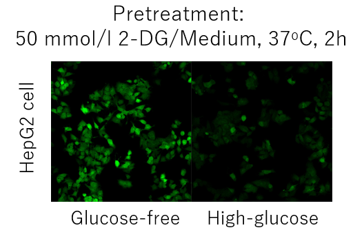
-
Q
Is Glucose Uptake Probe-Blue degraded or metabolized after uptaking by cells?
-
A
The structure of Glucose Uptake Probe-Blue is stable and will not be degraded during any steps of the protocol. The glucose moiety of Glucose Uptake Probe-Blue may undergo phosphorylation by hexokinase due to its structure, but no further metabolism is expected to occur.
-
Q
After Glucose Uptake Probe-Blue is uptaken by cells, can the cells be fixed?
-
A
No. Glucose Uptake Probe-Blue leaks from cells after fixation.
-
Q
Is that possible to store the working solution for a long time?
-
A
No, the Probe working solution cannot be stored. The Probe Stock solution can be stored at -20 oC for 1 month.
-
Q
What should I do if the sensitivity of measurement using Glucose Uptake Probe is low?
-
A
Please consider the probe concentration (×250 to ×1,000) and staining time (15 minutes to 1 hour) as initial considerations.
-
Q
What should I do if the fluorescence background is high in the measurement using Glucose Uptake Probe-Blue?
-
A
There is a possibility that some of the probes were not taken up by the cells remained. Please wash the cells again with WI solution.
-
Q
How long is the probe retained in the cells after washing with WI Solution?
-
A
The Probe is retained in the cells for about 1 hour at room temperature in WI Solution. Please note that the retention time may vary depending on the cell type.
-
Q
Can the Glucose Uptake Assay Kit-Blue be used to quantify glucose in the culture medium or in cells?
-
A
No, please use our product Glucose Assay Kit-WST (product code G264).
-
Q
Is it possible to quantify Glucose Uptake Probe taken up into cells?
-
A
No, this kit measures the fluorescence intensity of the probe taken up into the cell as an indication of the glucose uptake capacity of the cell.
Handling and storage condition
| 0-5°C |
Related products
The following related products are also used in research related to this product.


Product Classification
Product Classification
-
Cell Proliferation / Cell Cytotoxicity Assay
Cell Proliferation / Cell Cytotoxicity Assay Kits /Related Reagents
-
Cell Staining
Cell Double Staning Kit /Live Cell Staining /Dead Cell Staining /Nuclear Staining /Mitochondria Staning /Tissue Staining /Nucleolus Staining /Lipid Droplet Staining /Cell Membrane Staining /Lysosome Staining
-
Intracellular Fluorescent Probes
Reagents for Intracellular Calucuum Ion /Reagents for Intracellular Ion /Related Reagents
-
Labeling Chemistry
Protein Labeling Kits /Protein Labeling Reagents /HPLC Derivertization Reagents /Biotion Labeling Reagents /Related Reagents /Exsosome Labeling
-
Oxidative Stress
Stress Maker Detection /NO Detection /NO Donor /NO Inhibitor /ACE Inhibition Assay /Reagents・Kits for Sulfur Biology /Antioxidant Assay Kit /Donors for Sulfur Biology
-
-Bacstain- Series
Bacterial Proliferation Assay Kit /Bacteria Staining /Bacterial Fluorescent Staining
-
Molecular Biology
Transfection Reagents /Nuclear Staining /Agarose /Related Reagents /Buffer for Molecular Biology
-
Detergents
Detergents /Sets
-
Cross-Linking Reagents
Hetero-bifunctional Reagents /Homo-bifunctional Reagents /Others
-
Redox Dyes
Reductive Chromogenic Dyes /Electron Mediators /Oxidative Chromogenic Dyes /Trinder Reagents
-
Ion Analysis
Ionophores /Anion Eliminator /Solvent for Ion Electrode of Liquid Film Type
-
Buffers
Buffers
-
Metal Chelates
EDTA /Other Chelator /Reagents for Chelator Titration
-
Chromogen/Metal Indicator
Chromogen/Metal Indicator
-
Biochemicals
Biochemicals
-
Functional Organic Material
Alkanethiol Derivative /Phosphonic Acid Derivatives
