|
Senescence is a cellular process that results in the cessation of cell division, often serving as a protective mechanism against the proliferation of damaged cells, including potential cancer cells. This process is intricately regulated by numerous factors including, but not limited to, tumor suppressor genes, DNA damage response (DDR) pathways, and various signaling molecules. In addition, the senescence-associated secretory phenotype (SASP), consisting of cytokines, growth factors, and proteases, is regulated by NF-κB and other transcription factors that influence the tissue microenvironment and impact aging and disease processes. |
|||
|
HKDC1, a target of TFEB, is essential to maintain both mitochondrial and lysosomal homeostasis, preventing cellular senescence |
Iron accumulation drives fibrosis, senescence and the senescence-associated secretory phenotype Click here for the original article: Mate Maus, et. al., Nature, 2023. |
Microautophagy regulated by STK38 and GABARAPs is essential to repair lysosomes and prevent aging Click here for the original article: Monami Ogura, et. al., EMBP Reports, 2023. |
|
|
Point of Interest - This activity helps avert cellular senescence, playing a vital role in maintaining cellular homeostasis. - Beyond its role in glycolysis, HKDC1 contributes to mitophagy and lysosomal repair processes independently. - The absence of HKDC1 may result in cellular senescence and the buildup of damaged organelles.
|
Point of Interest - Senescent cells persistently accumulate iron, even after the increase in extracellular iron has subsided. - Cells exposed to various types of senescence-inducing insults accumulate abundant ferritin-bound iron, mostly within lysosomes. |
Point of Interest - Microautophagy in the repair of damaged lysosomes prevents aging. - STK38 and GABARAPs are key regulators of this process. - STK38 is required for lysosomal recruitment of VPS4 and GABARAPs are involved in ESCRT assembly. - Depletion of these regulators leads to accelerated cellular senescence and shortened lifespan. |
|
| Related Techniques | |||
| Cellular senescence detection | SPiDER-βGal for live-cell imaging or flow cytometry / microplate reader / tissue samples. | ||
| First-time autophagy research | Autophagic Flux Assay Kit | ||
| Autophagy detection | DAPGreen / DAPRed (Autophagosome detection), DALGreen (Autolysosome detection) | ||
| Lysosomal function | Lysosomal Acidic pH Detection Kit-Green/Red and Green/Deep Red | ||
| Ferrous ion (Fe2+) detection | FerroOrange(intracellular), Mito-FerroGreen(mitochondria) | ||
| Mitochondrial superoxide detection | MitoBright ROS Deep Red - Mitochondrial Superoxide Detection | ||
| Oxygen consumption rate assay | Extracellular OCR Plate Assay Kit | ||
| Related Applications | |||
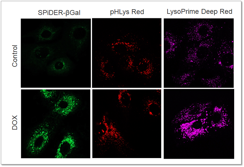
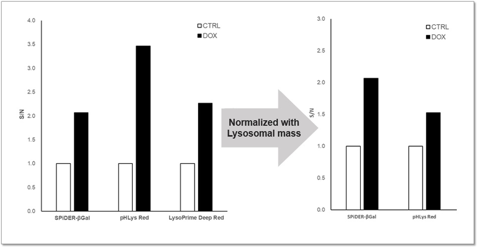
Analysis of Lysosomal Mass and pH change in Senescence-induced Cells |
|||
|
Purpose: To investigate changes in lysosomal mass and pH in A549 cells induced to senescence by treatment with Doxorubicin (DOX). Methods: Senescence-associated acidic β-galactosidase (SA-βGal) activity was detected using Cellular Senescence Detection Kit - SPiDER-βGal. Lysosomal mass was detected using LysoPrime Deep Red, and pH was detected using pHLys Red. Fluorescence imaging was used to observe changes in lysosomal mass and pH in senescent cells compared to non-senescent cells. The normalized fluorescence intensity of lysosomal mass and pH was also measured by a plate reader. Results: Our findings indicate that senescence induced by DOX resulted in an increase in lysosomal mass and acidification of pH compared to non-senescent cells. The obtained results are consistent with previous reports* that demonstrated enhanced lysosomal activity in senescent cells induced by the CDK4/6 inhibitor, palbociclib. The fluorescence imaging and plate reader data both support these findings. * Miguel Rovira, et. al., Aging Cell (2022) <Experimental Conditions for Microscopy> <Experimental Conditions for Plate Reader> <Products in Use> |
|
||