|
In recent years, the discovery of several novel senescence-related immune and neurological responses has attracted significant attention. The stress associated with mitochondrial protein folding is a driver of neural stem cell aging and suggests approaches to ameliorate aging-associated cognitive decline. In other breakthroughs, targeting bone marrow-derived senescent immune cells offers an avenue for improving brown adipose tissue aging and related metabolic disorders. Research also reveals the protective role of microglial autophagy in regulating the homeostasis of amyloid plaques and in preventing senescence; thus, the removal of senescent microglia emerges as a promising therapeutic strategy. |
||
|
The mitochondrial unfolded protein response regulates hippocampal neural stem cell aging |
Senescent immune cells accumulation promotes brown adipose tissue dysfunction during aging |
Autophagy enables microglia to engage amyloid plaques and prevents microglial senescence |
|
Point of Interest |
Point of Interest |
Point of Interest |
| Related Techniques | ||
| First choice for cellular senescence assay | Cellular Senescence Detection Kit – SPiDER-ßGal | |
| Cellular senescence assay with a plate reader | Cellular Senescence Plate Assay Kit – SPiDER-ßGal | |
| Lipid droplets detection | Lipi-Blue / Green / Red / Deep Red | |
| Fatty acid uptake assay | Fatty Acid Uptake Assay Kit | |
| Autophagy detection | DAPGreen / DAPRed (Autophagosome detection), DALGreen (Autolysosome detection) | |
| Mitochondrial function/glycolysis detection | Glycolysis/JC-1 MitoMP Assay Kit | |
| Oxygen consumption rate assay | Extracellular OCR Plate Assay Kit | |
| Related Applications | ||
|
|
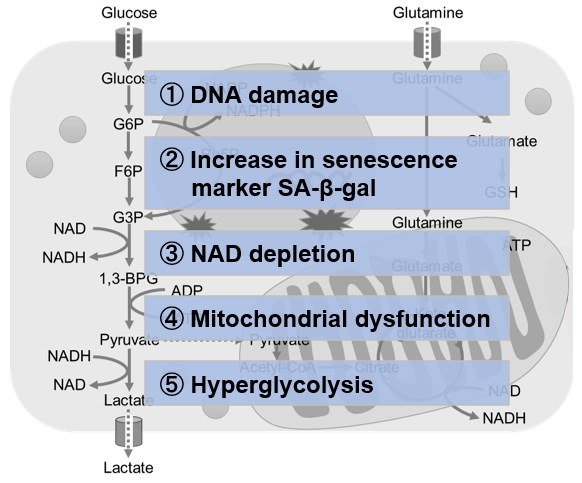
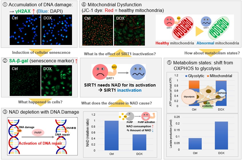
Metabolic shift to glycolysis in senescenct cellsNAD(+) levels decline during the aging process, causing defects in nuclear and mitochondrial functions and resulting in many age-associated pathologies*. Here, we try to redemonstrate this phenomenon in the doxorubicin (DOX)-induced cellular senescence model with a comprehensive analysis of our products. *S. Imai, et al., Trends Cell Biol, 2014, 24, 464-471
|
|
|
|
||