Controlling the immune cell helps treat cancer and neurodegenerative disease [Jul. 23, 2024]

[Jul. 23, 2024] Previous Science Note

Immune cell senescence is a critical factor in the progression of several diseases. As immune cells age and enter a senescent state, their ability to respond to infection, eliminate senescent cells, and regulate inflammation diminishes. This impaired immune function may contribute to the development and progression of age-related diseases, including cancer, cardiovascular disease, and neurodegenerative disorders. In addition, the accumulation of senescent immune cells in tissues can create a pro-inflammatory environment that further exacerbates disease pathology and impairs tissue function.

Senescence drives immunotherapy resistance by inducing an immunosuppressive tumor microenvironment
Click here for the original article: Damien Maggiorani, et. al., Nature Communications, 2024.

Point of Interest

- Immunotherapy resistance is associated with reduced CD8 T cell activity in tumors.

- Elimination of senescent cells restores CD8 T cell proliferation and reduces immunotherapy resistance.

- Anti-senescent cell drugs prior to immune checkpoint inhibitors may enhance the effectiveness of cancer immunotherapy.

Senescent alveolar macrophages promote early-stage lung tumorigenesis
Click here for the original article: Luis I. Prieto et. al., Cancer Cell, 2023.

Point of Interest

- Senescent alveolar macrophages accumulate early in lung cancer neoplasia and suppress cytotoxic T-cell responses.

- Removal of these macrophages reduces adenoma development, indicating their role in promoting tumorigenesis.

- Targeting senescent macrophages may slow lung cancer progression by altering the local microenvironment.

Rejuvenating aged microglia by p16ink4a-siRNA-loaded nanoparticles increases amyloid-β clearance in animal models of Alzheimer’s disease
Click here for the original article: Hyo Jung Shin et. al., Molecular Neurodegeneration, 2024.

Point of Interest

- Reduced amyloid clearance in Alzheimer's disease is associated with decreased phagocytic ability of older microglia.

- Downregulation of p16ink4a, a cell cycle factor associated with microglial aging, by siRNA increases amyloid clearance by phagocytosis and reverses cognitive deficits in Alzheimer's disease mode.

- Targeting p16ink4a with nanoparticles to rejuvenate microglia offers a potential treatment strategy for Alzheimer's disease.

Related Techniques

Endocytosis Detection detection
ECGreen-Endocytosis Detection
Lysosomal function
Lysosomal Acidic pH Detection Kit -Green/Red and Green/Deep Red
Cellular senescence detection
SPiDER-βGal for live-cell imaging or flow cytometry / microplate reader / tissue samples.
 
Plasma Membrane Staining
PlasMem Bright Green / Red
Ready-to-use kit for Cell cycle assay
Cell Cycle Assay Solution Blue / Deep Red
Total ROS detection
Highly sensitive DCFH-DA or Photo-oxidation Resistant DCFH-DA

Related Applications

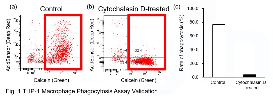
Phagocytosis assay of labeled apoptotic cells in THP-1 cells

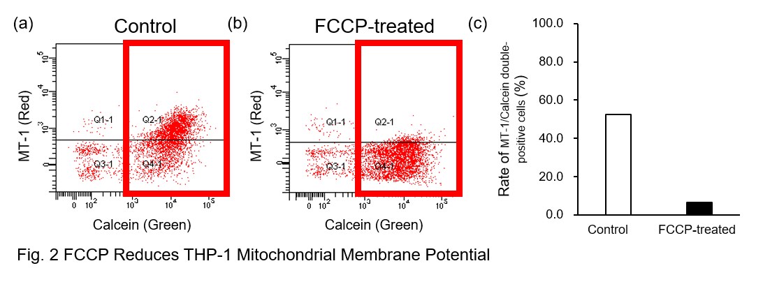
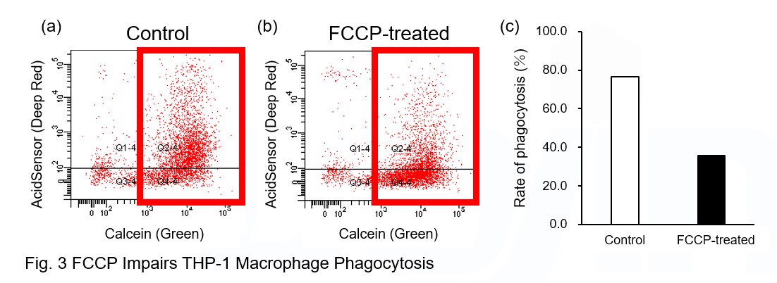
AcidSensor-labeled substances are taken up by cells and their fluorescence increases when they reach acidic organelles such as lysosomes. Taking advantage of this property, we evaluate the phagocytic activity of apoptotic cells by co-culturing AcidSensor-labeled apoptotic cells with Calcein-labeled THP-1 macrophages.  As a result, Calcein (Green) / AcidSensor (Deep red) double-positive cells, indicating THP-1 macrophages phagocytosing apoptotic cells, were observed by flow cytometry (Fig. 1a). Furthermore, when the phagocytosis of THP-1 macrophages was inhibited by Cytochalasin D, the percentage of double-positive cells decreased (Fig. 1b and 1c), confirming that the assay system can accurately evaluate phagocytosis.

A recent report reveals that inhibition of mitochondrial function induces a switch to glycolysis and reduces phagocytosis in cultured microglia, resident macrophages in the central nervous system*. To replicate this result, phagocytosis assays were performed using mitochondria-inhibited THP-1 macrophages. The results show that FCCP, a potent uncoupler of oxidative phosphorylation in mitochondria, decreases mitochondrial membrane potential (MT-1, Red) of THP-1 macrophages (Fig. 2) and reduces phagocytosis (Fig. 3).

*Lauren H. Fairley, et al., PNAS (2023)


Products in Use
① AcidSensor Labeling Kit – Endocytic Internalization Assay [code: A558]
② -Cellstain- Calcein-AM solution [code: C396]
 MT-1 MitoMP Detection Kit [code: MT13]

Product Classification

Product Classification