

|
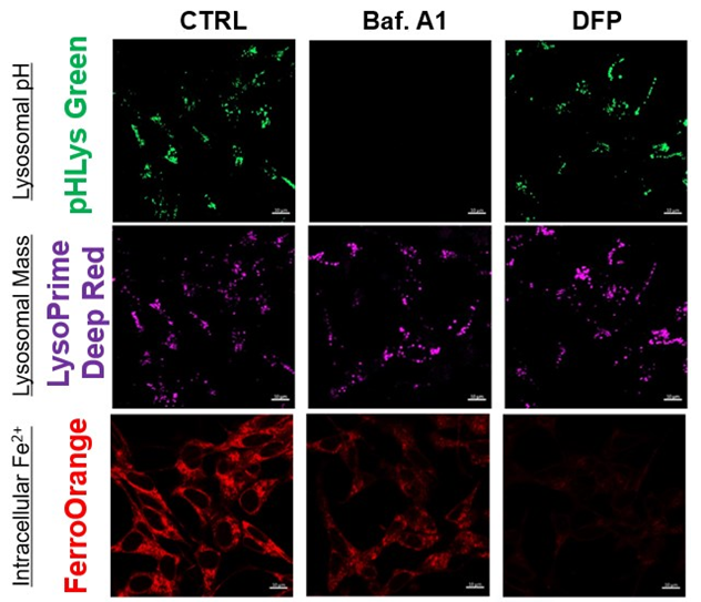
Scientists have unveiled that the selective autophagy adaptor NCOA4, which targets ferritin (a process known as ferritinophagy), is upregulated in pancreatic cancer. This upregulation ensures the availability of iron, thus encouraging tumor progression. Fascinatingly, ferritinophagy facilitates the synthesis of iron–sulfur cluster proteins, which help maintain mitochondrial homeostasis. Discover how the authors employed separate iron-detecting probes: FerroOrange for the cytosol and Mito-FerroGreen for the mitochondria (refer to Figure 1I and Supplemental Figure S4J). |
|
NCOA4-Mediated Ferritinophagy Is a Pancreatic Cancer Dependency via Maintenance of Iron Bioavailability for Iron–Sulfur Cluster Proteins Click here for the original article: Naiara Santana-Coodina, et. al., Cancer Discovery (2022) Point of Interest
|
| Related Techniques |
|
|
|
|
|
| Related Applications |
The simultaneous detection of lysosomal function with Mitochondrial ROS and intracellular Fe2+Lysosomal Function and Iron Homeostasis
|
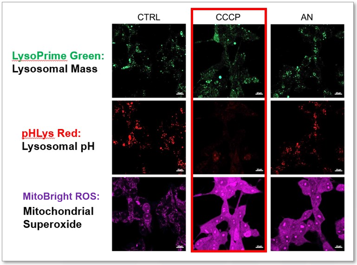
Lysosomal Function and Mitochondrial ROS
|


Product Classification
Product Classification
-
Cell Proliferation / Cell Cytotoxicity Assay
Cell Proliferation / Cell Cytotoxicity Assay Kits /Related Reagents
-
Cell Staining
Cell Double Staning Kit /Live Cell Staining /Dead Cell Staining /Nuclear Staining /Mitochondria Staning /Tissue Staining /Nucleolus Staining /Lipid Droplet Staining /Cell Membrane Staining /Lysosome Staining
-
Intracellular Fluorescent Probes
Reagents for Intracellular Calucuum Ion /Reagents for Intracellular Ion /Related Reagents
-
Labeling Chemistry
Protein Labeling Kits /Protein Labeling Reagents /HPLC Derivertization Reagents /Biotion Labeling Reagents /Related Reagents /Exsosome Labeling
-
Oxidative Stress
Stress Maker Detection /NO Detection /NO Donor /NO Inhibitor /ACE Inhibition Assay /Reagents・Kits for Sulfur Biology /Antioxidant Assay Kit /Donors for Sulfur Biology
-
-Bacstain- Series
Bacterial Proliferation Assay Kit /Bacteria Staining /Bacterial Fluorescent Staining
-
Molecular Biology
Transfection Reagents /Nuclear Staining /Agarose /Related Reagents /Buffer for Molecular Biology
-
Detergents
Detergents /Sets
-
Cross-Linking Reagents
Hetero-bifunctional Reagents /Homo-bifunctional Reagents /Others
-
Redox Dyes
Reductive Chromogenic Dyes /Electron Mediators /Oxidative Chromogenic Dyes /Trinder Reagents
-
Ion Analysis
Ionophores /Anion Eliminator /Solvent for Ion Electrode of Liquid Film Type
-
Organic Scintillator
-
Buffers
Buffers
-
Metal Chelates
EDTA /Other Chelator /Reagents for Chelator Titration
-
Chromogen/Metal Indicator
Chromogen/Metal Indicator
-
Water Analysis
/Fluorine /Iron / /Water Hardness /Residual Chlorine /ABS /Cyan / /Chromium /Copper
-
Extraction Reagent
AA Chelator /Related Reagents
-
High Purity Solvent
Spectrozole /Luminazole / /Acnazole /Dehydration Solvent for Synthesis
-
Biochemicals
Biochemicals
-
Functional Organic Material
Alkanethiol Derivative /Phosphonic Acid Derivatives
