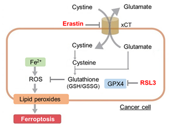
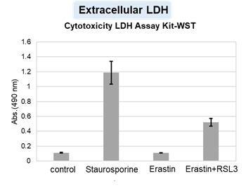
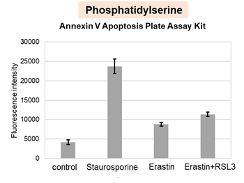
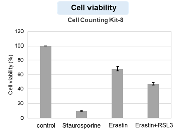
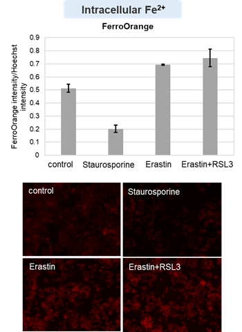
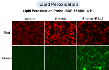
Hot Topic
Mechanisms and role of ferroptosis in disease
The field of ferroptosis research has grown exponentially in the past few years. This unique cell death by iron-dependent phospholipid peroxidation is regulated by multiple cellular metabolic pathways, including redox homeostasis, iron handling, mitochondrial activity, amino acid, lipid, and sugar metabolism, as well as various disease-related signaling pathways. Today, we introduce you to three highlighted articles focusing on iron resources, regulators, and the sensitive phenotype for ferroptosis in several diseases. |
The sensitive phenotype for ferroptosis |
Iron source in heart failure |
Regulator of ferroptosis |
Microglia ferroptosis is regulated by SEC24B and contributes to neurodegeneration
(Sean K. Ryan, et al., Nature Neuroscience, 26, 12-26, 2023) |
Iron derived from autophagy-mediated ferritin degradation induces cardiomyocyte death and heart failure in mice
(Jumpei Ito, et al., eLife, 10:e62174, 2021) |
The MARCHF6 E3 ubiquitin ligase acts as an NADPH sensor for the regulation of ferroptosis
(Kha The Nguyen, et al., Nature Cell Biology, 24, 1239-1251, 2022) |
- - iPS cell-derived tri-culture system that contains microglia, neurons, and astrocytes are used in this study
- - Microglia grown in a tri-culture system are highly responsive to iron and susceptible to ferroptosis
- - Iron overload causes a marked shift in the microglial transcriptional state
- - This microglial response contributes to neurodegeneration and is regulated by a novel ferroptosis susceptibility gene, SEC24B
|
- - Iron release from ferritin storage is through NCOA4-mediated autophagic degradation, known as ferritinophagy
- - Deletion of Ncoa4 in mouse hearts improved cardiac function along with the attenuation of the upregulation of ferritinophagy-mediated ferritin degradation 4 weeks after pressure overload
- - Free ferrous iron overload and increased lipid peroxidation were suppressed in NCOA4-deficient hearts
- - Inhibition of lipid peroxidation significantly mitigated the development of pressure overload-induced dilated cardiomyopathy in wild-type mice
|
- - The level of the anabolic reductant NADPH is a biomarker of ferroptosis sensitivity
- - The transmembrane endoplasmic reticulum MARCHF6 E3 ubiquitin ligase recognizes NADPH through its C-terminal regulatory region
- - This interaction upregulates the E3 ligase activity of MARCHF6, thus downregulating ferroptosis
- - Inhibiting ferroptosis rescued the growth of MARCHF6-deficient tumours and peri-natal lethality of Marchf6–/– mice.
|
Related Technique in This Topic |
Intracellular lipid peroxidation measurement |
Liperfluo HOT |
Mitochondria lipid peroxidation measurement |
MitoPeDPP |
Mitochondria ferrous ion (Fe2+) detection |
Mito-FerroGreen |
Intracellular ferrous ion (Fe2+) detection |
FerroOrange HOT |
Total ROS detection |
High Sensitive DCFH-DA HOT or Compatible with Immunostaining HOT |
Autophagy detection |
DAPGreen / DAPRed (Autophagosome detection), DALGreen (Autolysosome detection) |
GSSG/GSH assay |
GSSG/GSH Assay Kit |
Glutamine or Glutamate assay |
Glutamine Assay Kit, Glutamate Assay Kit |
NADP/NADPH assay |
NADP/NADPH Assay KIt |
Learn more about application data with multiple products here
Induction of Ferroptosis by Erastin?
Erastin is a known inducer of ferroptosis. By inhibiting the cystine transporter (xCT), erastin inhibits the uptake of cystine. Cystine is the raw material for GSH. Therefore, Erastin ultimately decreases the amount of GSH. Decreased GSH then results in lipid peroxide accumulation and induction of ferroptosis.
The following experimental examples show changes in each aforementioned index as a consequence of erastin stimulation. Measurements are made using Dojindo reagents.
Using erastin-treated A549 cells, we measured intracellular Fe2+, ROS, lipid peroxide, glutathione, glutamate release into the extracellular space, and cystine uptake. As a result, inhibition of xCT by elastin was observed and also the release of glutamate and uptake of cystine were decreased. Furthermore, elastin treatment decreased intracellular glutathione while it increased intracellular Fe2+ , ROS, and lipid peroxides.