

|
Metabolic alterations play a key role in determining the fate of cancer cells. Dysregulated metabolism, often characterized by increased glucose uptake and altered nutrient utilization, provides cancer cells with the necessary energy and building blocks necessary for rapid proliferation. In addition, metabolic reprogramming can confer resistance to apoptosis and promote tumor progression. Targeting these metabolic vulnerabilities holds promise for the development of therapeutic strategies that selectively eradicate cancer cells while sparing normal tissues. Understanding the intricate interplay between metabolism and cancer cell fate is essential for advancing personalized cancer therapies. |
|
| Related Techniques |
|
|
|
|
|
|
|
|
|
|
|
| Related Applications |
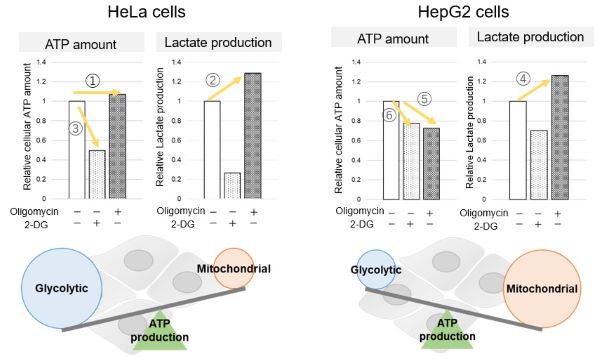
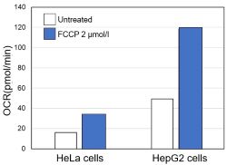
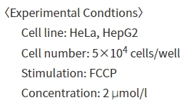
Comparison of metabolic pathway in two types of cancer cellsThe dependence of OXPHOS and Glycolysis in two types of cancer cells, HeLa and HepG2, were compared based on Lactate production, ATP levels, and OCR values. Many cancer cells produce ATP through the glycolytic pathway. On the other hand, it has been recently reported that cancer cells whose glycolytic pathway is suppressed survive by shifting their energy metabolism to OXPHOS by enhancing mitochondrial function, and the dependency of metabolic pathways differs depending on cell lines. |
<Evaluation by Lactate production and ATP levels>
|


Product Classification
Product Classification
-
Cell Proliferation / Cell Cytotoxicity Assay
Cell Proliferation / Cell Cytotoxicity Assay Kits /Related Reagents
-
Cell Staining
Cell Double Staning Kit /Live Cell Staining /Dead Cell Staining /Nuclear Staining /Mitochondria Staning /Tissue Staining /Nucleolus Staining /Lipid Droplet Staining /Cell Membrane Staining /Lysosome Staining
-
Intracellular Fluorescent Probes
Reagents for Intracellular Calucuum Ion /Reagents for Intracellular Ion /Related Reagents
-
Labeling Chemistry
Protein Labeling Kits /Protein Labeling Reagents /HPLC Derivertization Reagents /Biotion Labeling Reagents /Related Reagents /Exsosome Labeling
-
Oxidative Stress
Stress Maker Detection /NO Detection /NO Donor /NO Inhibitor /ACE Inhibition Assay /Reagents・Kits for Sulfur Biology /Antioxidant Assay Kit /Donors for Sulfur Biology
-
-Bacstain- Series
Bacterial Proliferation Assay Kit /Bacteria Staining /Bacterial Fluorescent Staining
-
Molecular Biology
Transfection Reagents /Nuclear Staining /Agarose /Related Reagents /Buffer for Molecular Biology
-
Detergents
Detergents /Sets
-
Cross-Linking Reagents
Hetero-bifunctional Reagents /Homo-bifunctional Reagents /Others
-
Redox Dyes
Reductive Chromogenic Dyes /Electron Mediators /Oxidative Chromogenic Dyes /Trinder Reagents
-
Ion Analysis
Ionophores /Anion Eliminator /Solvent for Ion Electrode of Liquid Film Type
-
Organic Scintillator
-
Buffers
Buffers
-
Metal Chelates
EDTA /Other Chelator /Reagents for Chelator Titration
-
Chromogen/Metal Indicator
Chromogen/Metal Indicator
-
Water Analysis
/Fluorine /Iron / /Water Hardness /Residual Chlorine /ABS /Cyan / /Chromium /Copper
-
Extraction Reagent
AA Chelator /Related Reagents
-
High Purity Solvent
Spectrozole /Luminazole / /Acnazole /Dehydration Solvent for Synthesis
-
Biochemicals
Biochemicals
-
Functional Organic Material
Alkanethiol Derivative /Phosphonic Acid Derivatives
