| [May. 9, 2023] | Previous Science Note |
|
Here, the scientists reveal an unexpected role for the lysosomal protein prosaposin (PSAP), the knockdown of which caused the formation of lipofuscin, a hallmark of aging, which traps iron, generating reactive oxygen species and triggering ferroptosis. Intriguingly, PSAP deficiency caused these dramatic phenotypes only in neurons, but not in other cells. Learn how the authors used Dojindo's Ferroptosis-related products, FerroOrange and Liperfluo for detecting Iron levels and lipid peroxidation, in their study. |
|
|
Related Techniques |
|
|
|
|
|
|
|
|
|
Related Applications |
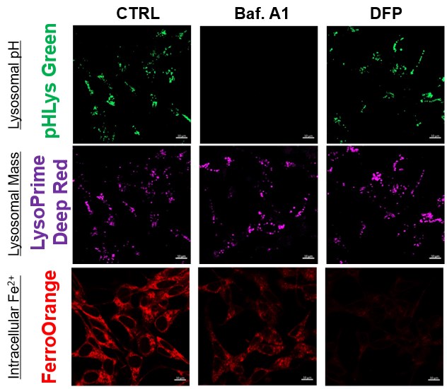
The simultaneous detection of lysosomal function with intracellular Fe2+
Recent reports suggest that lysosomal neutralization can result in iron depletion, consequently leading to the disruption of cell viability. To verify this, HeLa cells were labeled with FerroOrange for Fe2+ detection, and the lysosomal mass and pH were separately detected with LysoPrime DeepRed and pHLys Green (a product currently under development). Co-staining with FerroOrange and Lysosomal dyes demonstrated that Bafilomycin A1 (Baf. A1), an inhibitor of lysosomal acidification, causes iron depletion consistent with the findings reported in the article. Interestingly, the iron chelator, Deferiprone (DFP), did not impact lysosomal pH, suggesting that lysosomal function plays a key role in managing iron homeostasis. Reference: Ross A Weber, et. al., Mol Cell (2020) Products in Use *pHLys Green is available as the "Lysosomal Acidic pH Detection Kit-Green/Deep Red". |