| Target |
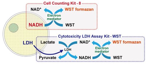
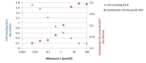
Released Lacate dehydrogenase (LDH) |
A marker (Calcein-AM) released into the supernatant (Calcein-Release Assay) |
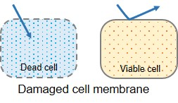
Cytoplasm(Live cell membranes are impermeable to PI, but not dead cell membranes) |
Cytoplasm; cells taking up Trypan Blue are considered non-viable |
| Instrument |
Microplate Reader
(colorimetric) |
Microplate reader, FCM, Fluorescence microscope
(Fluorometric) |
Fluorescence microscope, FCM |
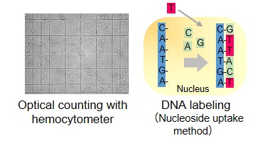
Microscope
(colorimetric) |
| Advantage |
·Simple procedure
·Suitable for multi-sample assay |
·Convenient for cytotoxicity assay using 2+ kinds of cell samples |
·Low price
·Simple procedure |
·Low price
·No special instruments needed |
| Disadvantage |
·Increased background by serum |
·Leakage of reagent from living cells |
·Not applicable for multi-sample assay |
·False-positives due to cytotoxicity
·Not applicable for multi-sample assay |
| Product code |
CK12 |
CK06 (kit)
C396 (ready-to-use solution)
C326 (powder) |
P378 (ready-to-use solution)
P346 (powder) |
– |


 Please watch this video for comparison between MTT and Cell Counting Kit-8.