


Science Note: Senescence
Senescence Driven by Mitochondrial Alterations [Mar. 5, 2025]
Open / Close the Article
| Cellular senescence is closely linked to mitochondrial dysfunction, which drives aging and disease through metabolic shifts, oxidative stress, and pro-inflammatory signaling. This Science Note shows the latest findings on mitochondrial mechanisms that promote cellular senescence. | ||||||||||||||||||||||||
|
Apoptotic stress causes mtDNA release during senescence and drives the SASP Highlighted technique: When apoptotic stress increases the permeability of the mitochondrial outer membrane, cytochrome c is released into the cytoplasm, making it a key indicator of MOMP. In this study, MOMP is assessed by co-staining the mitochondrial outer membrane protein TOM20 and cytochrome c, followed by high-resolution analysis. |
||||||||||||||||||||||||
|
Mitochondrial fatty acid oxidation drives senescence Highlighted technique: This study assessed senescence using multiple markers, including p16 expression and SA-βgal activity. While other markers changed, γH2AX did not, reinforcing the widely accepted notion that no single marker defines senescence. Thus, multiple indicators are needed to assess senescence. |
||||||||||||||||||||||||
|
Senescent glia link mitochondrial dysfunction and lipid accumulation |
||||||||||||||||||||||||
Related Techniques (click to open/close)
|
||||||||||||||||||||||||
Application Note (click to open/close)
|
||||||||||||||||||||||||
|
|
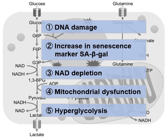
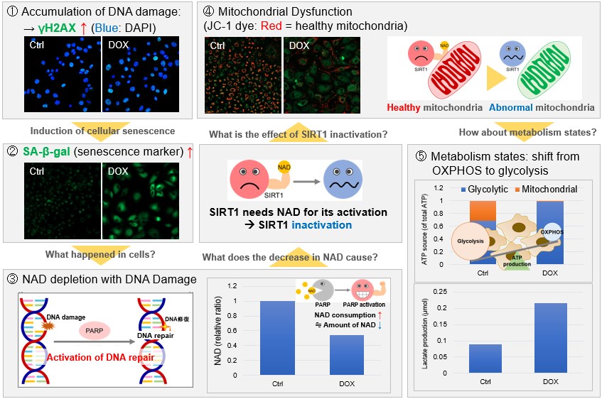
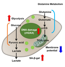
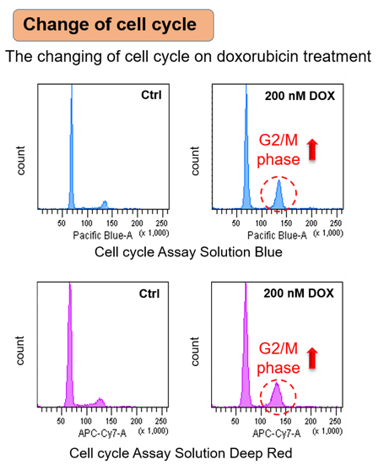
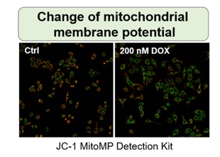
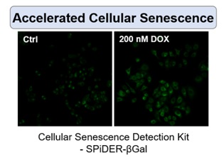
NAD(+) levels decline during the aging process, causing defects in nuclear and mitochondrial functions and resulting in many age-associated pathologies*. Here, we try to redemonstrate this phenomenon in the doxorubicin (DOX)-induced cellular senescence model with a comprehensive analysis of our products. *S. Imai, et al., Trends Cell Biol, 2014, 24, 464-471
|
|
|
|

Are Interleukin Therapies the Future of Anti-Aging? [Jan. 28, 2025]
Open / Close the Article
|
Chronic inflammation drives immune decline and age-related diseases, creating a vicious cycle with senescence. This week, we introduce a review of the link between senescence and inflammation, along with recent discoveries. Cellular senescence and inflammation are linked, as senescent cells secrete pro-inflammatory factors that perpetuate chronic inflammation and tissue dysfunction. Chronic inflammation drives immune decline and age-related diseases, creating a vicious cycle with senescence. In this note, we introduce a review article of the link between senescence and inflammation, along with recent findings on anti-IL-17 and anti-IL-11 treatment for anti-senescence. Anti-IL-17 drugs are already approved, and anti-IL-11 therapy is in trials for fibrotic lung disease, suggesting that these treatments may soon help delay aging. |
|||
|
Review Article Inflammation and aging: signaling pathways and intervention therapies |
Inhibition of IL-11 signalling extends mammalian healthspan and lifespan |
Targeting lymphoid-derived IL-17 signaling to delay skin aging Click here for the original article: Paloma Solá, et. al., Nature Aging, 2024. |
|
|
Point of Interest - Inflammaging creates a vicious cycle of inflammation and senescence, suggesting the elimination of inflammation as a potential anti-aging strategy. - The paper reviews inflammaging, aging models, single-cell technologies, and anti-aging strategies to combat disease and improve quality of life. |
Point of Interest - Genetic deletion of IL-11 or use of anti-IL-11 improves metabolism, reduces frailty and extends lifespan by over 20% in mice. - Anti-IL-11 therapy, currently in clinical trials for fibroinflammatory diseases, may extend health and lifespan in humans with promising safety. |
Point of Interest - Single-cell RNA sequencing reveals increased IL-17 signaling in aged skin, which drives inflammation and impairs homeostasis. - Blocking IL-17 signaling reduces skin inflammation and delays age-related features, suggesting a potential anti-aging skin therapy. |
|
| Related Techniques | |||
| Cellular senescence detection | SPiDER-βGal for live-cell imaging or flow cytometry / microplate reader / tissue samples NEW SPiDER-βGal Blue for fixed cell and for multiple staining with immunostaining and other methods |
||
| Oxygen Consumption Rate(OCR) Plate Assay | Extracellular OCR Plate Assay Kit | ||
| Total ROS detection | Highly sensitive DCFH-DA or Photo-oxidation Resistant DCFH-DA | ||
| Glycolysis/Oxidative phosphorylation Assay | Glycolysis/OXPHOS Assay Kit | ||
| First-time autophagy research | Autophagic Flux Assay Kit | ||
| Lysosomal function | Lysosomal Acidic pH Detection Kit-Green/Red and Green/Deep Red | ||
| Apoptosis detection in multiple samples | Annexin V Apoptosis Plate Assay Kit | ||
| Cell proliferation/ cytotoxicity assay | Cell Counting Kit-8 and Cytotoxicity LDH Assay Kit-WST | ||
| Related Applications | |||
Metabolic shift to glycolysis in senescenct cells |
|||
|
|
NAD(+) levels decline during the aging process, causing defects in nuclear and mitochondrial functions and resulting in many age-associated pathologies*. Here, we try to redemonstrate this phenomenon in the doxorubicin (DOX)-induced cellular senescence model with a comprehensive analysis of our products. *S. Imai, et al., Trends Cell Biol, 2014, 24, 464-471
|
||
 |
|||
Multiple staining with oxidative stress-related markers using Doxorubicin-induced senescent cells(flow cytometry) |
|||
|
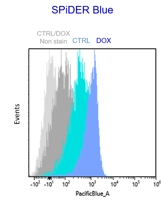
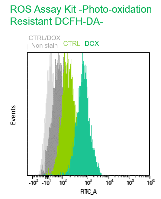
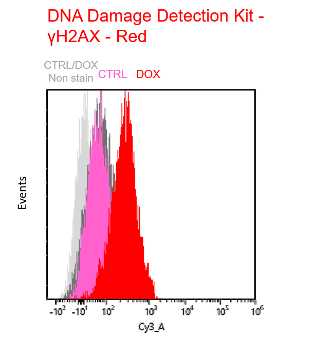
Using A549 cells induced to senescence by doxorubicin (DOX) and normal cells (CTRL), changes in oxidative stress-related markers in senescent cells were analyzed by flow cytometry with multiple staining. SA-βGal as a senescence marker was detected by Cellular Senescence Detection Kit - SPiDER Blue, total ROS as an oxidative stress marker was detected by ROS Assay Kit - Photo-oxidation Resistant DCFH-DA-, and γH2AX as a DNA damage marker was detected by DNA Damage Detection Kit - γH2AX-Red. As a result, total ROS and γH2AX were increased in SA-βGal-positive senescent cells, and the increase in oxidative stress-related markers associated with cellular senescence could be detected by multiple staining. <Experimental Procedure> |
|||


Senescent Cells Foster a Tumor-Promoting Microenvironment [Jan. 7, 2025]
Open / Close the Article
|
Recent research on cellular senescence reveals that senescent cells contribute to tumor promotion. Here are some papers that highlight how senescent cells create a pro-tumorigenic microenvironment by enhancing inflammation, angiogenesis, and immune evasion, offering therapeutic opportunities. Cellular senescence is a state of stable cell cycle arrest that prevents the proliferation of damaged or aged cells and acts as a tumor suppressor mechanism. However, senescent cells can secrete a pro-inflammatory and tissue-altering mix of factors that can paradoxically promote cancer progression. Senescent factors contribute to a pro-tumorigenic microenvironment by enhancing inflammation, angiogenesis, and immune evasion. Understanding the dual role of senescence in cancer highlights the complexity of its contribution to tumor suppression and progression and offers potential therapeutic opportunities. |
|||
|
Hematopoietic aging promotes cancer by fueling IL-1⍺–driven emergency myelopoiesis |
Age-dependent loss of HAPLN1 erodes vascular integrity via indirect upregulation of endothelial ICAM1 in melanoma |
Fibroblasts in the Aged Pancreas Drive Pancreatic Cancer Progression Click here for the original article: Daniel J. Zabransky, et. al., Cancer Research, 2024. |
|
|
Point of Interest - DNMT3A (DNA methyltransferase 3A) downregulation with age increases IL-1⍺ production, driving the recruitment of immunosuppressive myeloid cells and tumor progression. - Blocking the IL-1 receptor slows lung cancer progression in aged mice, highlighting potential therapeutic strategies for age-related cancers. |
Point of Interest - ICAM1 leads to VE-cadherin internalization, which increases vascular permeability and melanoma progression. - Blocking ICAM1 reduces tumor size and metastasis in aged mice, highlighting the impact of aging and ECM changes on tumor progression.
|
Point of Interest - Treatment of young mice with GDF-15 enhances tumor growth, whereas GDF-15 knockout in aged mice reduces tumor growth. - AKT inhibition is effective in aged but not young microenvironments, providing a targeted therapy for age-related pancreatic cancer. |
|
| Related Techniques | |||
| Cellular senescence detection | SPiDER-βGal for live-cell imaging or flow cytometry / microplate reader / tissue samples SPiDER-βGal Blue for fixed cell and for multiple staining with immunostaining and other methods |
||
| Total ROS detection | Highly sensitive DCFH-DA or Photo-oxidation Resistant DCFH-DA | ||
| Glycolysis/Oxidative phosphorylation Assay | Glycolysis/OXPHOS Assay Kit | ||
| Oxygen Consumption Rate(OCR) Plate Assa | Extracellular OCR Plate Assay Kit | ||
| First-time autophagy research | Autophagic Flux Assay Kit | ||
| Lysosomal function | Lysosomal Acidic pH Detection Kit-Green/Red and Green/Deep Red | ||
| Apoptosis detection in multiple samples | Annexin V Apoptosis Plate Assay Kit | ||
| Cell proliferation/ cytotoxicity assay | Cell Counting Kit-8 and Cytotoxicity LDH Assay Kit-WST | ||
| Related Applications | |||
Metabolic shift to glycolysis in senescenct cells |
|||
|
|
NAD(+) levels decline during the aging process, causing defects in nuclear and mitochondrial functions and resulting in many age-associated pathologies*. Here, we try to redemonstrate this phenomenon in the doxorubicin (DOX)-induced cellular senescence model with a comprehensive analysis of our products. *S. Imai, et al., Trends Cell Biol, 2014, 24, 464-471
|
||
|
|||
Multiple staining with oxidative stress-related markers using Doxorubicin-induced senescent cells(flow cytometry) |
|||
|
Using A549 cells induced to senescence by doxorubicin (DOX) and normal cells (CTRL), changes in oxidative stress-related markers in senescent cells were analyzed by flow cytometry with multiple staining. SA-βGal as a senescence marker was detected by Cellular Senescence Detection Kit - SPiDER Blue, total ROS as an oxidative stress marker was detected by ROS Assay Kit - Photo-oxidation Resistant DCFH-DA-, and γH2AX as a DNA damage marker was detected by DNA Damage Detection Kit - γH2AX-Red. As a result, total ROS and γH2AX were increased in SA-βGal-positive senescent cells, and the increase in oxidative stress-related markers associated with cellular senescence could be detected by multiple staining. <Experimental Procedure> |
|||
The Spread of Senescent Cells: From Local to Systemic [Dec. 10, 2024]
Open / Close the Article
|
Recent research on cellular senescence shows that senescent cells propagate the effects of aging and contribute to chronic inflammation and diseases such as neurodegeneration. Here are studies that explore the systemic effects of senescence and therapeutic strategies that target senescent cells and their secreted factors. Senescent cells are dysfunctional cells that accumulate in organs and tissues with age and secrete bioactive factors, collectively known as the senescence-associated secretory phenotype (SASP). These factors influence the local microenvironment and can propagate senescence to nearby and distant cells, leading to systemic effects. Systemic senescence contributes to age-related tissue dysfunction, chronic inflammation and the progression of diseases such as cancer, cardiovascular disease and neurodegeneration. Targeting senescent cells or their secreted factors offers a promising therapeutic strategy to attenuate systemic aging and related pathologies. |
|||
|
Hepatocellular senescence induces multi-organ senescence and dysfunction via TGFβ |
Aged bone marrow macrophages drive systemic aging and age-related dysfunction via extracellular vesicle-mediated induction of paracrine senescence |
Senescent cell transplantation into the skin induces age-related peripheral dysfunction and cognitive decline Click here for the original article: Ana Catarina Franco, et. al., Aging Cell, 2024. |
|
|
Point of Interest - Hepatocyte senescence predicts the outcome of acute liver failure, including organ failure, driven by the systemic effects of the TGFβ signaling pathway. - Blocking TGFβ inhibits the spread of senescence and prevents liver-induced kidney dysfunction, highlighting the role of senescence in multi-organ dysfunction. |
Point of Interest - The PPARα agonist fenofibrate restores tissue homeostasis in aged mice and reduces the risk of chronic diseases, suggesting a potential to extend human lifespan. - BMMs are key to senescence propagation, linking aging dysfunction to a treatable mechanism with therapeutic implications.
|
Point of Interest - The senescent fibroblasts in the skin increase mobility and musculoskeletal impairment, demonstrating systemic effects of skin-resident senescence. - Skin senescence correlates with hippocampal dysfunction, linking local senescence to brain dysfunction and age-related cognitive decline. |
|
| Related Techniques | |||
| Cellular senescence detection | SPiDER-βGal for live-cell imaging or flow cytometry / microplate reader / tissue samples NEW SPiDER-βGal Blue for fixed cell and for multiple staining with immunostaining and other methods |
||
| Total ROS detection | Highly sensitive DCFH-DA or Photo-oxidation Resistant DCFH-DA | ||
| Glycolysis/Oxidative phosphorylation Assay | Glycolysis/OXPHOS Assay Kit | ||
| Oxygen Consumption Rate(OCR) Plate Assa | Extracellular OCR Plate Assay Kit | ||
| First-time autophagy research | Autophagic Flux Assay Kit | ||
| Lysosomal function | Lysosomal Acidic pH Detection Kit-Green/Red and Green/Deep Red | ||
| Lipid Droplet detection | Lipid Droplet Assay Kit - Blue / Deep Red | ||
| Exosome staining | ExoSparkler Exosome Membrane Labeling Kit-Green / Red / Deep Red | ||
| Apoptosis detection in multiple samples | Annexin V Apoptosis Plate Assay Kit | ||
| Cell proliferation/ cytotoxicity assay | Cell Counting Kit-8 and Cytotoxicity LDH Assay Kit-WST | ||
| Related Applications | |||
Co-staining with Lipid droplet and SA-β-Gal in fixed cells |
|||
|
|
NAD(+) levels decline during the aging process, causing defects in nuclear and mitochondrial functions and resulting in many age-associated pathologies*. Here, we try to redemonstrate this phenomenon in the doxorubicin (DOX)-induced cellular senescence model with a comprehensive analysis of our products. *S. Imai, et al., Trends Cell Biol, 2014, 24, 464-471
|
||
 |
|||
Multiple staining with oxidative stress-related markers using Doxorubicin-induced senescent cells(flow cytometry) |
|||
|
Using A549 cells induced to senescence by doxorubicin (DOX) and normal cells (CTRL), changes in oxidative stress-related markers in senescent cells were analyzed by flow cytometry with multiple staining. SA-βGal as a senescence marker was detected by Cellular Senescence Detection Kit - SPiDER Blue, total ROS as an oxidative stress marker was detected by ROS Assay Kit - Photo-oxidation Resistant DCFH-DA-, and γH2AX as a DNA damage marker was detected by DNA Damage Detection Kit - γH2AX-Red. As a result, total ROS and γH2AX were increased in SA-βGal-positive senescent cells, and the increase in oxidative stress-related markers associated with cellular senescence could be detected by multiple staining. <Experimental Procedure> |
|||
Targeting Senescent Cells to Enhance Cancer Treatment Outcomes [Oct. 22, 2024]
Open / Close the Article
|
Recent cancer research has focused on intratumoral senescent cancer cells and it is becoming clear that these cells contribute to immune evasion of cancer cells and create a cancer-promoting environment. Here are some of the papers that have identified senescent cells or senescent cells with specific phenotypes in cancer treatment and have shown that removing these cells is important for cancer treatment. Senescent cells are damaged or stressed cells that permanently stop dividing but do not undergo cell death. In the context of cancer, senescent cells can accumulate in tumors, particularly after treatments such as chemotherapy, contributing to an immunosuppressive environment. This environment can promote tumor growth, immune evasion and resistance to therapy. However, the selective targeting and elimination of senescent cells, known as senolytic therapy, is emerging as a promising strategy to improve cancer treatment by reducing tumor progression and improving patient outcomes. |
|||
|
The efficacy of chemotherapy is limited by intratumoral senescent cells expressing PD-L2 |
Cellular senescence in malignant cells promotes tumor progression in mouse and patient Glioblastoma |
Senescence drives immunotherapy resistance by inducing an immunosuppressive tumor microenvironment Click here for the original article: Damien Maggiorani, et. al., Nature Communications, 2024. |
|
|
Point of Interest - PD-L2 is not necessary for senescence but crucial for immune evasion by senescent cells, promoting tumor persistence after chemotherapy. - Antibody-mediated PD-L2 blockade synergizes with chemotherapy, suggesting a potential therapeutic strategy targeting senescent cancer cells. |
Point of Interest - Removal of p16Ink4a-expressing senescent cells from GBM tumors improves survival in mice and alters the tumour environment. - Senolytic drugs that target senescent cells may be a promising adjuvant therapy for GBM patients, improving survival. |
Point of Interest - Elimination of senescent cells restores CD8 T cell proliferation and reduces immunotherapy resistance. - Anti-senescent cell drugs prior to immune checkpoint inhibitors may enhance the effectiveness of cancer immunotherapy. |
|
| Related Techniques | |||
| Cellular senescence detection | SPiDER-βGal for live-cell imaging or flow cytometry / microplate reader / tissue samples NEW SPiDER-βGal Blue for fixed cell and for multiple staining with immunostaining and other methods |
||
| Glycolysis/Oxidative phosphorylation Assay | Glycolysis/OXPHOS Assay Kit | ||
| Endocytosis Detection detection | ECGreen-Endocytosis Detection | ||
| Lysosomal function | Lysosomal Acidic pH Detection Kit-Green/Red and Green/Deep Red | ||
| First-time autophagy research | Autophagic Flux Assay Kit | ||
| Total ROS detection | Highly sensitive DCFH-DA or Photo-oxidation Resistant DCFH-DA | ||
| Apoptosis detection in multiple samples | Annexin V Apoptosis Plate Assay Kit | ||
| Cell proliferation/ cytotoxicity assay | Cell Counting Kit-8 and Cytotoxicity LDH Assay Kit-WST | ||
| Glutathione Quantification | GSSG/GSH Quantification Kit | ||
| Related Applications | |||
Co-staining with Lipid droplet and SA-β-Gal in fixed cells |
|||
|
|
NAD(+) levels decline during the aging process, causing defects in nuclear and mitochondrial functions and resulting in many age-associated pathologies*. Here, we try to redemonstrate this phenomenon in the doxorubicin (DOX)-induced cellular senescence model with a comprehensive analysis of our products. *S. Imai, et al., Trends Cell Biol, 2014, 24, 464-471
|
||
|
|||
Multiple staining with oxidative stress-related markers using Doxorubicin-induced senescent cells(flow cytometry) |
|||
|
Using A549 cells induced to senescence by doxorubicin (DOX) and normal cells (CTRL), changes in oxidative stress-related markers in senescent cells were analyzed by flow cytometry with multiple staining. SA-βGal as a senescence marker was detected by Cellular Senescence Detection Kit - SPiDER Blue, total ROS as an oxidative stress marker was detected by ROS Assay Kit - Photo-oxidation Resistant DCFH-DA-, and γH2AX as a DNA damage marker was detected by DNA Damage Detection Kit - γH2AX-Red. As a result, total ROS and γH2AX were increased in SA-βGal-positive senescent cells, and the increase in oxidative stress-related markers associated with cellular senescence could be detected by multiple staining. <Experimental Procedure> |
|||
Senescence and Lipid Droplet Accumulation are Involved in the Onset of Neurodegenerative Diseases [Sep. 10, 2024]
Open / Close the Article
|
Recent research on senescence is revealing that senescence is related to lipid droplet and lysosomal dysfunction, which impair cell function. Here are some of the papers showing that these processes leads to neurodegenerative diseases. Cellular senescence, a state of irreversible growth arrest, is closely linked to neurodegeneration through the accumulation of damaged cells in the nervous system. Lipid droplets, which store excess lipids, can accumulate in aging or stressed cells, contributing to cellular dysfunction and exacerbating neurodegenerative processes. Lysosomal dysfunction plays a central role in both lipid accumulation and the removal of cellular waste, and its impairment leads to the accumulation of toxic substances that further drive neurodegeneration. Together, these mechanisms create a cycle of cellular damage that accelerates the progression of neurodegenerative diseases such as Alzheimer's and Parkinson's. |
|||
| Lipid accumulation drives cellular senescence in dopaminergic neurons Click here for the original article: Taylor Russo, et. al., Aging, 2024. |
Senescent glia link mitochondrial dysfunction and lipid accumulation Click here for the original article: China Byrns, et. al., Nature, 2024.
|
BHLHE40/41 regulate microglia and peripheral macrophage responses associated with Alzheimer’s disease and other disorders of lipid-rich tissues Click here for the original article: Anna Podleśny-Drabiniok, et. al., Nature Communications, 2024. |
|
|
Point of Interest - Mutations in the lysosomal enzyme β-glucocerebrosidase cause lipid accumulation that drives cellular senescence in dopaminergic neurons in PD. - Lipid droplet aggregation and lysosomal dysfunction may trigger cellular senescence leading to neurodegeneration in PD. |
Point of Interest - AP1+ senescent glia cause lipid accumulation in non-senescent glia and increase senescence markers. - Targeting AP1 in senescent glia extends lifespan, but increases oxidative damage in the brain and neuronal mitochondrial function remains poor. |
Point of Interest - DLAMs are macrophage subpopulations with similar transcriptional activation states found in aging brains and other diseased lipid-rich tissues. - Targeting BHLHE40/41, transcriptional regulators of DALM, may improve cholesterol clearance and lysosomal function in Alzheimer's disease therapies.
|
|
| Related Techniques | |||
| Cellular senescence detection | SPiDER-βGal for live-cell imaging or flow cytometry / microplate reader / tissue samples NEW SPiDER-βGal Blue for fixed cell and for multiple staining with immunostaining and other methods |
||
| Lipid Droplet detection | Lipid Droplet Assay Kit - Blue / Deep Red | ||
| Lipid Droplet Staining | Lipi-Blue/ Green/ Red/ Deep Red | ||
| Lysosomal function | Lysosomal Acidic pH Detection Kit-Green/Red and Green/Deep Red | ||
| First-time autophagy research | Autophagic Flux Assay Kit | ||
| Mitochondrial membrane potential detection | JC-1 MitoMP Detection Kit, MT-1 MitoMP Detection Kit | ||
| Mitochondrial superoxide detection | MitoBright ROS Deep Red - Mitochondrial Superoxide Detection | ||
| Glycolysis/Oxidative phosphorylation Assay | Glycolysis/OXPHOS Assay Kit | ||
| Glutathione Quantification | GSSG/GSH Quantification Kit | ||
 |
|||
| Related Applications | |||
Co-staining with Lipid droplet and SA-β-Gal in fixed cells |
|||
|
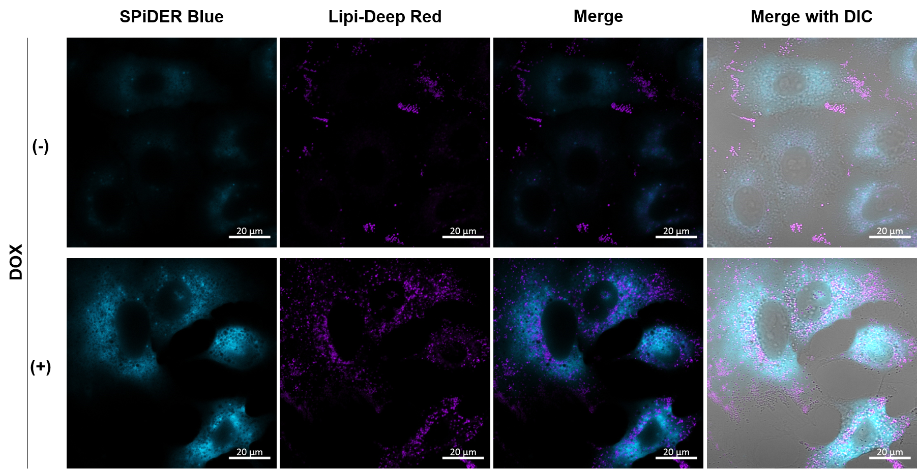
1. A549 (2 x 10⁴) cells were seeded onto µ-slide 8 well plates (ibidi) and cultured overnight in a 37°C CO2 incubator.
|
Imaging analysis of lipid droplet accumulation in senescent cells was performed using normal A549 cells (CTRL) and cells induced senescence by Doxorubicin treatment (DOX). SA-β-Gal was detected as a senescence marker with Cellular Senescence Detection Kit - SPiDER Blue, and lipid droplets were detected with Lipi-Deep Red. As a result, the signal of Lipi-Deep Red was increased in SA-β-Gal-positive senescent cells.
[Detection conditions] SPiDE Blue: 405 nm (Ex), 400–550 nm (Em), 1.0%, 600V
|
||
Multiple staining with oxidative stress-related markers using Doxorubicin-induced senescent cells(flow cytometry) |
|||
|
Using A549 cells induced to senescence by doxorubicin (DOX) and normal cells (CTRL), changes in oxidative stress-related markers in senescent cells were analyzed by flow cytometry with multiple staining. SA-βGal as a senescence marker was detected by Cellular Senescence Detection Kit - SPiDER Blue, total ROS as an oxidative stress marker was detected by ROS Assay Kit - Photo-oxidation Resistant DCFH-DA-, and γH2AX as a DNA damage marker was detected by DNA Damage Detection Kit - γH2AX-Red. As a result, total ROS and γH2AX were increased in SA-βGal-positive senescent cells, and the increase in oxidative stress-related markers associated with cellular senescence could be detected by multiple staining. <Experimental Procedure> |
|||
Anti-Senescence Targets Based on Metabolic Pathways [Aug. 27, 2024]
Open / Close the Article
|
Anti-senescence strategies often target metabolic pathways to delay or reverse cellular aging processes. Key pathways such as the pentose phosphate pathway (PPP) and mitochondrial metabolism are modulated to reduce oxidative stress and increase cellular resilience. By optimizing these pathways, anti-senescence approaches aim to maintain energy production, prevent accumulation of damage, and preserve stem cell function. Ultimately, these interventions may improve tissue homeostasis and mitigate age-related decline. |
|||
| A homoeostatic switch causing glycerol-3-phosphate and phosphoethanolamine accumulation triggers senescence by rewiring lipid metabolism Click here for the original article: Khaled Tighanimine, et. al., Nature Metabolism, 2024. |
Proline restores mitochondrial function and reverses aging hallmarks in senescent cells Click here for the original article: Debanik Choudhury, et. al., Cell Reports, 2024.
|
Citrate metabolism controls the senescent microenvironment via the remodeling of pro-inflammatory enhancers Click here for the original article: Kan Etoh, et. al., Cell Reports, 2024. |
|
|
Point of Interest - p53-dependent glycerol kinase activation and post-translational modifications drive this metabolic switch, promoting triglyceride accumulation and senescence. - Scavenging G3P and pEtN reduces their accumulation, acting senomorphically and potentially providing a therapeutic approach to target senescence. |
Point of Interest - Proline supplementation induces mitophagy, alleviates mitochondrial respiratory impairment and improves mitochondrial clearance in senescent cells. - Proline treatment mitigates the hallmarks of aging, including DNA damage, inflammation, and impaired differentiation, by restoring mitochondrial function. |
Point of Interest - ACLY-dependent acetyl-CoA activates SASP gene enhancers, increasing BRD4 recruitment and promoting SASP activation. - Chemical inhibition of the ACLY-BRD4 axis suppresses SASP in aged mice, providing a potential target for promoting healthy aging.
|
|
| Related Techniques | |||
| Cellular senescence detection | SPiDER-βGal for live-cell imaging or flow cytometry / microplate reader / tissue samples. | ||
| Mitochondrial membrane potential detection | JC-1 MitoMP Detection Kit, MT-1 MitoMP Detection Kit | ||
| First-time autophagy research | Autophagic Flux Assay Kit | ||
| Lysosomal function | Lysosomal Acidic pH Detection Kit-Green/Red and Green/Deep Red | ||
| Glycolysis/Oxidative phosphorylation Assay | Glycolysis/OXPHOS Assay Kit | ||
| Oxygen Consumption Rate(OCR) Plate Assay | Extracellular OCR Plate Assay Kit | ||
| Lipid Droplet detection | Lipid Droplet Assay Kit - Blue / Deep Red | ||
| Glutathione Quantification | GSSG/GSH Quantification Kit | ||
| Total ROS detection | Highly sensitive DCFH-DA or Photo-oxidation Resistant DCFH-DA | ||
| Endocytosis Detection detection | ECGreen-Endocytosis Detection | ||
| Related Applications | |||
Metabolic shift to glycolysis in senescenct cells |
|||
|
|
NAD(+) levels decline during the aging process, causing defects in nuclear and mitochondrial functions and resulting in many age-associated pathologies*. Here, we try to redemonstrate this phenomenon in the doxorubicin (DOX)-induced cellular senescence model with a comprehensive analysis of our products. *S. Imai, et al., Trends Cell Biol, 2014, 24, 464-471
|
||
|
|||
Anti-Senescence Effects Induced by Extracellular Vesicles [Jul. 2, 2024]
Open / Close the Article
|
Anti-senescence study aims to restore youthful function to aging tissues and organs, often focusing on cellular repair and regeneration. Extracellular vesicles (EVs), including exosomes and microvesicles, play a critical role in cell-to-cell communication by transporting bioactive molecules such as proteins, lipids and RNA. Recent studies suggest that EVs derived from young or healthy cells can have anti-senescence effects on aged cells and tissues, promoting regeneration and repair. These EVs have shown potential to reverse age-related damage and improve tissue function, making them a promising tool in regenerative medicine and anti-aging therapies. |
|||
|
Small extracellular vesicles from young plasma reverse age-related functional declines by improving mitochondrial energy metabolism |
Extracellular vesicles from human urine-derived stem cells delay aging through the transfer of PLAU and TIMP1 Click here for the original article: Shanshan Rao, et. al., Acta Biomaterialia, 2024. |
Rejuvenating effects of young extracellular vesicles in aged rats and in cellular models of human senescence Click here for the original article: Lilian Grigorian Shamagian, et. al., EMBP Reports, 2023. |
|
|
Point of Interest - Proteomic analysis shows young sEVs enhance metabolism and mitochondrial function in aged tissues. - Young sEVs stimulate PGC-1α, reversing age-related dysfunction.
|
Point of Interest - USC-EVs' anti-aging effects are consistent regardless of donor age, gender, or health. - Plasminogen activator urokinase (PLAU) and tissue inhibitor of metalloproteinases 1 (TIMP1) in USC-EVs contribute to the anti-senescent effects of USC-EVs. |
Point of Interest - Young cardiosphere-derived cells (CDC) EVs rejuvenate aged rats, improving heart, lungs, muscles, and kidneys. - Purified EVs from young blood show rejuvenating effects; EV-depleted serum increases senescence. - The treatment also favorably modulates glucose metabolism and anti-senescence pathways, ultimately extending lifespan. |
|
| Related Techniques | |||
| Cellular senescence detection | SPiDER-βGal for live-cell imaging or flow cytometry / microplate reader / tissue samples. | ||
| Exosome Labeling | ExoSparkler Exosome Membrane Labeling Kit - Green / Red / Deep Red | ||
| Exosome purification | ExoIsolator Exosome Isolation Kit, ExoIsolator Isolation Filter | ||
| Glycolysis/Oxidative phosphorylation Assay | Glycolysis/OXPHOS Assay Kit | ||
| Oxygen Consumption Rate(OCR) Plate Assay | Extracellular OCR Plate Assay Kit | ||
| Mitochondrial membrane potential detection | JC-1 MitoMP Detection Kit, MT-1 MitoMP Detection Kit | ||
| Mitochondrial superoxide detection | MitoBright ROS Deep Red - Mitochondrial Superoxide Detection | ||
| Total ROS detection | Highly sensitive DCFH-DA or Photo-oxidation Resistant DCFH-DA | ||
| Endocytosis Detection detection | ECGreen-Endocytosis Detection | ||
| Lysosomal function | Lysosomal Acidic pH Detection Kit-Green/Red and Green/Deep Red | ||
| Related Applications | |||
Metabolic shift to glycolysis in senescenct cells |
|||
|
|
NAD(+) levels decline during the aging process, causing defects in nuclear and mitochondrial functions and resulting in many age-associated pathologies*. Here, we try to redemonstrate this phenomenon in the doxorubicin (DOX)-induced cellular senescence model with a comprehensive analysis of our products. *S. Imai, et al., Trends Cell Biol, 2014, 24, 464-471
|
||
|
|||
Mitochondrial and Lysosomal, and Iron Regulation of Senescence [Mar. 26, 2024]
Open / Close the Article
|
Senescence is a cellular process that results in the cessation of cell division, often serving as a protective mechanism against the proliferation of damaged cells, including potential cancer cells. This process is intricately regulated by numerous factors including, but not limited to, tumor suppressor genes, DNA damage response (DDR) pathways, and various signaling molecules. In addition, the senescence-associated secretory phenotype (SASP), consisting of cytokines, growth factors, and proteases, is regulated by NF-κB and other transcription factors that influence the tissue microenvironment and impact aging and disease processes. |
|||
|
HKDC1, a target of TFEB, is essential to maintain both mitochondrial and lysosomal homeostasis, preventing cellular senescence |
Iron accumulation drives fibrosis, senescence and the senescence-associated secretory phenotype Click here for the original article: Mate Maus, et. al., Nature, 2023. |
Microautophagy regulated by STK38 and GABARAPs is essential to repair lysosomes and prevent aging Click here for the original article: Monami Ogura, et. al., EMBP Reports, 2023. |
|
|
Point of Interest - This activity helps avert cellular senescence, playing a vital role in maintaining cellular homeostasis. - Beyond its role in glycolysis, HKDC1 contributes to mitophagy and lysosomal repair processes independently. - The absence of HKDC1 may result in cellular senescence and the buildup of damaged organelles.
|
Point of Interest - Senescent cells persistently accumulate iron, even after the increase in extracellular iron has subsided. - Cells exposed to various types of senescence-inducing insults accumulate abundant ferritin-bound iron, mostly within lysosomes. |
Point of Interest - Microautophagy in the repair of damaged lysosomes prevents aging. - STK38 and GABARAPs are key regulators of this process. - STK38 is required for lysosomal recruitment of VPS4 and GABARAPs are involved in ESCRT assembly. - Depletion of these regulators leads to accelerated cellular senescence and shortened lifespan. |
|
| Related Techniques | |||
| Cellular senescence detection | SPiDER-βGal for live-cell imaging or flow cytometry / microplate reader / tissue samples. | ||
| First-time autophagy research | Autophagic Flux Assay Kit | ||
| Autophagy detection | DAPGreen / DAPRed (Autophagosome detection), DALGreen (Autolysosome detection) | ||
| Lysosomal function | Lysosomal Acidic pH Detection Kit-Green/Red and Green/Deep Red | ||
| Ferrous ion (Fe2+) detection | FerroOrange(intracellular), Mito-FerroGreen(mitochondria) | ||
| Mitochondrial superoxide detection | MitoBright ROS Deep Red - Mitochondrial Superoxide Detection | ||
| Oxygen consumption rate assay | Extracellular OCR Plate Assay Kit | ||
| Related Applications | |||
Analysis of Lysosomal Mass and pH change in Senescence-induced Cells |
|||
|
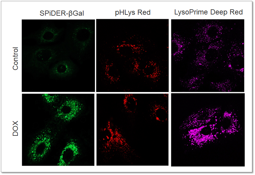
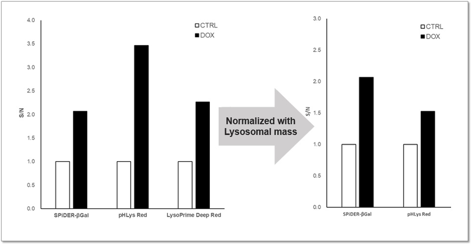
Purpose: To investigate changes in lysosomal mass and pH in A549 cells induced to senescence by treatment with Doxorubicin (DOX). Methods: Senescence-associated acidic β-galactosidase (SA-βGal) activity was detected using Cellular Senescence Detection Kit - SPiDER-βGal. Lysosomal mass was detected using LysoPrime Deep Red, and pH was detected using pHLys Red. Fluorescence imaging was used to observe changes in lysosomal mass and pH in senescent cells compared to non-senescent cells. The normalized fluorescence intensity of lysosomal mass and pH was also measured by a plate reader. Results: Our findings indicate that senescence induced by DOX resulted in an increase in lysosomal mass and acidification of pH compared to non-senescent cells. The obtained results are consistent with previous reports* that demonstrated enhanced lysosomal activity in senescent cells induced by the CDK4/6 inhibitor, palbociclib. The fluorescence imaging and plate reader data both support these findings. * Miguel Rovira, et. al., Aging Cell (2022) <Experimental Conditions for Microscopy> <Experimental Conditions for Plate Reader> <Products in Use> |
|
||
Senescence Related to Reprogramming and Regeneration [Sep. 26, 2023]
Open / Close the Article
|
Scientists have discovered that, in the absence of resident stem cells, senescent cells can instruct neighboring somatic cells to reprogram. This reprogramming enables them to become stem cells, driving whole-body regeneration in the cnidarian Hydractinia symbiolongicarpus. |
|||
|
Senescence-induced cellular reprogramming drives cnidarian whole-body regeneration |
|||
|
Point of Interest |
|||
| Related Techniques | |||
| First choice for cellular senescence assay | Cellular Senescence Detection Kit – SPiDER-ßGal | ||
| Cellular senescence assay with a plate reader | Cellular Senescence Plate Assay Kit – SPiDER-ßGal | ||
| Mitophagy detection | Mitophagy Detection Kit and Mtphagy Dye | ||
| Cell cycle assay | Cell Cycle Assay Solution Blue / Deep Red | ||
| Dead cell staining for flow cytometry | Dead Cell Makeup Blue / Deep Red - Higher Retention than PI | ||
| Lysosomal pH detection | Lysosomal Acidic pH Detection Kit-Green/Red, Green/Deep Red | ||
| Mitochondrial function/glycolysis detection | Glycolysis/JC-1 MitoMP Assay Kit | ||
| Oxygen consumption rate assay | Extracellular OCR Plate Assay Kit | ||
| Related Applications | |||
| Regulating Cell Cycle Arrest: p16, p21, p53, and pRB | |||
|
Irreversible cell cycle arrest is one of the phenomena that characterize cellular senescence. p16, p21, p53, and pRB (phosphorylated retinoblastoma protein) are known as representative protein markers. The activation/upregulation of these proteins are used as indicators of cellular senescence. These marker proteins are known to be tumor suppressors and regulate the cell cycle mainly through two pathways (p16Ink4a-RB and p53-p21CIP1).
|
||
Dysregulated DNA Hydroxymethylation in Dyslipidemia-Related Apoptosis and Senescence [Oct. 03, 2023]
Open / Close the Article
|
Scientists have discovered that obesity and dyslipidemia are linked to dysregulated DNA hydroxymethylation in apoptosis- and senescence-related genes in both swine and human MSCs. This dysregulation might impact cell vitality and regenerative capabilities. Vitamin C could mediate the reprogramming of this altered epigenomic profile, offering a potential approach to enhance the success of autologous MSC transplantation in obese patients. |
|||
|
Obesity and dyslipidemia are associated with partially reversible modifications to DNA hydroxymethylation of apoptosis- and senescence-related genes in swine adipose-derived mesenchymal stem/stromal cells |
|||
|
Point of Interest |
|||
| Related Techniques | |||
| First choice for cellular senescence assay | Cellular Senescence Detection Kit – SPiDER-ßGal | ||
| Cellular senescence assay with a plate reader | Cellular Senescence Plate Assay Kit – SPiDER-ßGal | ||
| Mitophagy detection | Mitophagy Detection Kit and Mtphagy Dye | ||
| Lysosomal pH detection | Lysosomal Acidic pH Detection Kit-Green/Red, Green/Deep Red | ||
| Mitochondrial function/glycolysis detection | Glycolysis/JC-1 MitoMP Assay Kit | ||
| Oxygen consumption rate assay | Extracellular OCR Plate Assay Kit | ||
| Related Applications | |||
Metabolic shift to glycolysis in senescenct cells |
|||
 |
NAD(+) levels decline during the aging process, causing defects in nuclear and mitochondrial functions and resulting in many age-associated pathologies*. Here, we try to redemonstrate this phenomenon in the doxorubicin (DOX)-induced cellular senescence model with a comprehensive analysis of our products. *S. Imai, et al., Trends Cell Biol, 2014, 24, 464-471
|
||
 |
|||
Abrogated CBFb activity induces senescence-like phenotypes in macrophages [Sep. 19, 2023]
Open / Close the Article
|
Through single-cell RNA sequencing (scRNA-seq), they identified an indispensable transcription factor, CBFb that ensures AM self-renewal. The study concludes that AMs in aged hosts exhibit signs of senescence, possibly due to reduced activity of the CBFb transcription factor. |
|||
|
Single cell RNA sequencing unravels mechanisms underlying senescence-like phenotypes of alveolar macrophages |
|||
|
Point of Interest |
|||
| Related Techniques | |||
| First choice for cellular senescence assay | Cellular Senescence Detection Kit – SPiDER-ßGal | ||
| Cellular senescence assay with a plate reader | Cellular Senescence Plate Assay Kit – SPiDER-ßGal | ||
| Cell cycle assay | Cell Cycle Assay Solution Blue / Deep Red | ||
| Dead cell staining for flow cytometry | Dead Cell Makeup Blue / Deep Red - Higher Retention than PI | ||
| Lysosomal pH detection | Lysosomal Acidic pH Detection Kit-Green/Red, Green/Deep Red | ||
| Mitochondrial function/glycolysis detection | Glycolysis/JC-1 MitoMP Assay Kit | ||
| Oxygen consumption rate assay | Extracellular OCR Plate Assay Kit | ||
| Related Applications | |||
| Analysis of Lysosomal Mass and pH Exchange in Senescence-induced Cells | |||
|
|
Purpose: To investigate changes in lysosomal mass and pH in A549 cells induced to senescence by treatment with Doxorubicin (DOX). Methods: Senescence-associated acidic β-galactosidase (SA-βGal) activity was detected using Cellular Senescence Detection Kit - SPiDER-βGal. Lysosomal mass was detected using LysoPrime Deep Red, and pH was detected using pHLys Red. Fluorescence imaging was used to observe changes in lysosomal mass and pH in senescent cells compared to non-senescent cells. The normalized fluorescence intensity of lysosomal mass and pH was also measured by a plate reader. Results: Our findings indicate that senescence induced by DOX resulted in an increase in lysosomal mass and acidification of pH compared to non-senescent cells. The obtained results are consistent with previous reports* that demonstrated enhanced lysosomal activity in senescent cells induced by the CDK4/6 inhibitor, palbociclib. The fluorescence imaging and plate reader data both support these findings. * Miguel Rovira, et. al., Aging Cell (2022)
<Experimental Conditions for Microscopy> <Experimental Conditions for Plate Reader>
<Products in Use> |
||
MYC degradation-related cell cycle arrest in senescent cells [Sep. 14, 2023]
Open / Close the Article
|
Scientists have unveiled that senescence is irreversible and that commitment to and maintenance of senescence is mediated by irreversible MYC degradation. Thus, irreversible cell cycle exit associated with senescence is mediated by constitutive MYC degradation, but bypassing this degradation may allow tumor cells to escape during cancer initiation. |
|||
|
Irreversible cell cycle exit associated with senescence is mediated by constitutive MYC degradation |
|||
|
Point of Interest |
|||
| Related Techniques | |||
| First choice for cellular senescence assay | Cellular Senescence Detection Kit – SPiDER-ßGal | ||
| Cellular senescence assay with a plate reader | Cellular Senescence Plate Assay Kit – SPiDER-ßGal | ||
| Cell cycle assay | Cell Cycle Assay Solution Blue / Deep Red | ||
| Lysosomal pH detection | Lysosomal Acidic pH Detection Kit-Green/Red, Green/Deep Red | ||
| Mitochondrial function/glycolysis detection | Glycolysis/JC-1 MitoMP Assay Kit | ||
| Oxygen consumption rate assay | Extracellular OCR Plate Assay Kit | ||
| Related Applications | |||
|
|
Metabolic shift to glycolysis in senescenct cellsNAD(+) levels decline during the aging process, causing defects in nuclear and mitochondrial functions and resulting in many age-associated pathologies*. Here, we try to redemonstrate this phenomenon in the doxorubicin (DOX)-induced cellular senescence model with a comprehensive analysis of our products. *S. Imai, et al., Trends Cell Biol, 2014, 24, 464-471
|
||
|
|
|||
Senescence-related Stress Responses in Immuno and Neurological Aging [Aug 15, 2023]
Open / Close the Article
|
In recent years, the discovery of several novel senescence-related immune and neurological responses has attracted significant attention. The stress associated with mitochondrial protein folding is a driver of neural stem cell aging and suggests approaches to ameliorate aging-associated cognitive decline. In other breakthroughs, targeting bone marrow-derived senescent immune cells offers an avenue for improving brown adipose tissue aging and related metabolic disorders. Research also reveals the protective role of microglial autophagy in regulating the homeostasis of amyloid plaques and in preventing senescence; thus, the removal of senescent microglia emerges as a promising therapeutic strategy. |
||
|
The mitochondrial unfolded protein response regulates hippocampal neural stem cell aging |
Senescent immune cells accumulation promotes brown adipose tissue dysfunction during aging |
Autophagy enables microglia to engage amyloid plaques and prevents microglial senescence |
|
Point of Interest |
Point of Interest |
Point of Interest |
| Related Techniques | ||
| First choice for cellular senescence assay | Cellular Senescence Detection Kit – SPiDER-ßGal | |
| Cellular senescence assay with a plate reader | Cellular Senescence Plate Assay Kit – SPiDER-ßGal | |
| Lipid droplets detection | Lipi-Blue / Green / Red / Deep Red | |
| Fatty acid uptake assay | Fatty Acid Uptake Assay Kit | |
| Autophagy detection | DAPGreen / DAPRed (Autophagosome detection), DALGreen (Autolysosome detection) | |
| Mitochondrial function/glycolysis detection | Glycolysis/JC-1 MitoMP Assay Kit | |
| Oxygen consumption rate assay | Extracellular OCR Plate Assay Kit | |
| Related Applications | ||
|
|
Metabolic shift to glycolysis in senescenct cellsNAD(+) levels decline during the aging process, causing defects in nuclear and mitochondrial functions and resulting in many age-associated pathologies*. Here, we try to redemonstrate this phenomenon in the doxorubicin (DOX)-induced cellular senescence model with a comprehensive analysis of our products. *S. Imai, et al., Trends Cell Biol, 2014, 24, 464-471
|
|
|
|
||
Senescent Cells and Lysosomal Cholesterol Accumulation [Mar. 28, 2023]
Open / Close the Article
| This article focusing on the role of cholesterol accumulation in lysosomes of senescent cells, and its connection to maintaining the senescence-associated secretory phenotype (SASP). We believe that these findings can provide valuable insights into the relationship between senescence and lysosomal function. | |
|
Lysosomal control of senescence and inflammation through cholesterol partitioning Point of Interest |
|
| Related Techniques | |
| Cellular senescence detection (Live cell imaging or FCM) | Cellular Senescence Detection Kit HOT |
| Cellular senescence detection (Plate reader) | Cellular Senescence Plate Assay Kit HOT |
| Autophagy detection | DAPGreen / DAPRed (Autophagosome detection), DALGreen (Autolysosome detection) |
| Lysosomal function assay | Lysosomal pH and mass detection Kit |
| Lysosome staining | pH-dependent (Red) and pH-independent (Green / Deep Red) probes |
| Nutrient uptake Assay | Glucose (Blue / Green / Red), Amino Acid, Cystine, and Fatty Acid Uptake Assay Kit |
| Lipid droplets detection | Lipi-Blue / Green / Red / Deep Red |
| Related Applications | |
| Analysis of Lysosomal Mass and pH Exchange in Senescence-induced Cells | |
|
|
Purpose: To investigate changes in lysosomal mass and pH in A549 cells induced to senescence by treatment with Doxorubicin (DOX). Methods: Senescence-associated acidic β-galactosidase (SA-βGal) activity was detected using Cellular Senescence Detection Kit - SPiDER-βGal. Lysosomal mass was detected using LysoPrime Deep Red, and pH was detected using pHLys Red. Fluorescence imaging was used to observe changes in lysosomal mass and pH in senescent cells compared to non-senescent cells. The normalized fluorescence intensity of lysosomal mass and pH was also measured by a plate reader. Results: Our findings indicate that senescence induced by DOX resulted in an increase in lysosomal mass and acidification of pH compared to non-senescent cells. The obtained results are consistent with previous reports* that demonstrated enhanced lysosomal activity in senescent cells induced by the CDK4/6 inhibitor, palbociclib. The fluorescence imaging and plate reader data both support these findings. * Miguel Rovira, et. al., Aging Cell (2022)
<Experimental Conditions for Microscopy> <Experimental Conditions for Plate Reader>
<Products in Use> |

Reverse Electron Transfer in Age-Related Diseases [Mar. 21, 2023]
Open / Close the Article
| This article focusing on reverse electron transfer (RET) at mitochondrial complex I, which trigers age-related disease. These findings may help to understand the relationship between senescence and metabolism through mitochondrial dysfunction. | |||||
| Reverse electron transfer is activated during aging and contributes to aging and age-related
Suman Rimal, et. al., EMBO Reports, e55548 (2023) Point of Interest |
|||||
| Related Techniques | |||||
| Cellular senescence detection (Live cell imaging or FCM) | Cellular Senescence Detection Kit HOT | ||||
| Cellular senescence detection (Plate reader) | Cellular Senescence Plate Assay Kit HOT | ||||
| Oxygen consumption rate assay | Extracellular OCR Plate Assay Kit | ||||
| NAD/NADH assay | NAD/NADH Assay Kit | ||||
| Mitochondrial membrane potential detection | JC-1 MitoMP Detection Kit / MT-1 MitoMP Detection Kit | ||||
| Mitochondrial superoxide detection | MitoBright ROS Deep Red - Mitochondrial Superoxide Detection | ||||
| Total ROS detection | Higher sensitivity or Compatible with Co-staining for Immunostaining HOT | ||||
| Related Applications | |||||
Metabolic shift to glycolysis in senescenct cells
|
|||||

Diverse Functions of Senescent Cells [Feb. 14, 2023]
Open / Close the Article
| Cellular senescence, which contributes significantly to aging, is controlled by various factors such as cell type and physiological conditions, such as oxidative stress. It has been reported that these senescent cells have various negative effects, and treatment methods aimed at removing these senescent cells (Senotherapeutics) have been attracting attention. On the other hand, there are also reports of positive effects, and more detailed studies on the functions of senescent cells will be needed in the future. Today, we introduce you to three highlighted articles related to cellular senescence focusing on Senotherapeutics, Positive and negative impacts on tissue regeneration. | ||
|---|---|---|
| Senotherapeutics in Alzheimer's disease | Negative impacts on tissue regeneration | Positive impacts on tissue regeneration |
| Increased post-mitotic senescence in aged human neurons is a pathological feature of Alzheimer’s disease (Joseph R. Herdy, et al., Cell Stem Cell, 29, 1637-1652, 2022) |
Senescence atlas reveals an aged-like inflamed niche that blunts muscle regeneration (Victoria Moiseeva, et al., Nature, 613, 169-178, 2023) |
Sentinel p16INK4a+ cells in the basement membrane form a reparative niche in the lung (Nabora S. Reyes, et al., Science, 378, 192-201, 2022) |
|
|
|
| Related Technique in This Topic | ||
| Cellular senescence detection (Live cell imaging or FCM) |
Cellular Senescence Detection Kit - SPiDER-βGal | |
| Cellular senescence detection (Plate reader) | Cellular Senescence Plate Assay Kit - SPiDER-βGal HOT | |
| Oxygen consumption rate assay | Extracellular OCR Plate Assay Kit NEW | |
| Glycolysis/Oxidative phosphorylation Assay | Glycolysis/OXPHOS Assay Kit HOT | |
| NAD/NADH assay | NAD/NADH Assay Kit | |
| Mitochondrial membrane potential detection | JC-1 MitoMP Detection Kit and MT-1 MitoMP Detection Kit | |
| Mitochondrial superoxide detection | MitoBright ROS Deep Red - Mitochondrial Superoxide Detection HOT | |
| Total ROS detection | Higher sensitivity HOT or compatible with co-staining for immunostaining NEW | |
Learn more about application data with multiple products here
The Drivers of Cellular Senescence [Jan. 16, 2023]
Open / Close the Article
|
Cellular senescence is a complex biological process that is influenced by several key factors: DNA damage, telomere shortening, oxidative stress, oncogene activation, and so on. These factors collectively contribute to the complex process of cellular senescence, which acts as a double-edged sword - protective in preventing cancer growth, but also contributing to aging and age-related diseases. |
|||
|
Apoptotic stress causes mtDNA release during senescence and drives the SASP |
Iron accumulation drives fibrosis, senescence and the senescence-associated secretory phenotype Click here for the original article: Mate Maus, et. al., Nature, 2023. |
Genome-wide CRISPR activation screening in senescent cells reveals SOX5 as a driver and therapeutic target of rejuvenation Click here for the original article: Yaobin Jing, et. al., Cell Stem Cell, 2023. |
|
|
Point of Interest |
Point of Interest - Vascular and hemolytic injury trigger iron accumulation, which causes senescence and promotes fibrosis. - Senescent cells persistently accumulate iron, even after the increase in extracellular iron has subsided. - Cells exposed to various types of senescence-inducing insults accumulate abundant ferritin-bound iron, mostly within lysosomes. - The high levels of labile iron fuel the generation of reactive oxygen species and the SASP. |
Point of Interest - CRISPRa screening identifies a comprehensive set of rejuvenators against senescence. - Activation of SOX5 initiates a rejuvenation program via epigenetic remodeling. - SOX5 activation leads to the stimulation of the HMGB2 enhancer, resulting in subsequent geroprotective effects. - Gene therapy using only SOX5 has the potential to promote the regeneration of aged knee joints. |
|
| Related Techniques | |||
| Cellular senescence detection | SPiDER-βGal for live-cell imaging or flow cytometry / microplate reader / tissue samples. | ||
| Ferrous ion (Fe2+) detection | FerroOrange and Mito-FerroGreen | ||
| Total ROS detection | Highly sensitive DCFH-DA or Photo-oxidation Resistant DCFH-DA | ||
| Lysosomal function | Lysosomal Acidic pH Detection Kit-Green/Red and Green/Deep Red | ||
| Mitochondrial superoxide detection | MitoBright ROS Deep Red - Mitochondrial Superoxide Detection | ||
| Mitochondrial membrane potential detection | JC-1 MitoMP Detection Kit / MT-1 MitoMP Detection Kit | ||
| Oxygen consumption rate assay | Extracellular OCR Plate Assay Kit | ||
| Antibody/Protein labeling: quick and high recovery | Fluorescein, Biotin, and Peroxidase Labeling Kit - NH2 | ||
| Related Applications | |||
Metabolic shift to glycolysis in senescenct cells |
|||
| |
NAD(+) levels decline during the aging process, causing defects in nuclear and mitochondrial functions and resulting in many age-associated pathologies*. Here, we try to redemonstrate this phenomenon in the doxorubicin (DOX)-induced cellular senescence model with a comprehensive analysis of our products. *S. Imai, et al., Trends Cell Biol, 2014, 24, 464-471
|
||
|
|||
