


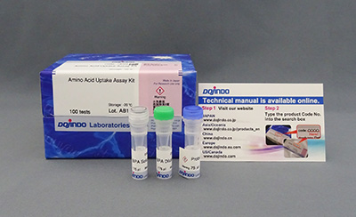
Amino Acid Uptake Assay Kit
Amino Acid Uptake Assay
-
Product codeUP04 Amino Acid Uptake Assay Kit
| Unit size | Price | FUJIFILM Wako Products code |
|---|---|---|
| 20 tests | ¥17,100 | 346-09891 |
| 100 tests | ¥48,200 | 342-09893 |
 The type of microplate you choose can significantly influence your results. Not all microplates are compatible with this assay.
Please refer to the FAQ for recommended microplates and their influence on measurement results.
| 20 tests | BPA Solution BPA Dilution Buffer Probe Solution |
35 µl×1 35 µl×1 15 µl×1 |
|---|---|---|
| 100 tests | BPA Solution BPA Dilution Buffer Probe Solution |
175 µl×1 175 µl×1 75 µl×1 |

Description
Amino acids are essential for intracellular protein and nucleic acid synthesis, Especially for cancer cells which are proliferating continually. Since the supply of acetyl CoA from the glycolytic pathway is decreased in cancer cells, they have a furthermore huge demand for amino acids which are an important nutrient source for the TCA cycle. The research has shown that cancer cells increase the expression of the amino acid transporter LAT1 (L-type amino acid transporter 1) and take up a large number of amino acids, which is expected to be a new target for anti-Cancer drugs' discovery.
In immunotherapy, not only the metabolic changes in cancer cells but also in immune cells are considered to affect the treatment. For example, as senescence occurs in immune cells, their metabolic system changes, and their immune capacity to attack cancer cells decreases. Therefore, research is being conducted on nutrient uptake regulation by immune cells and improving the effectiveness of cancer immunotherapy.
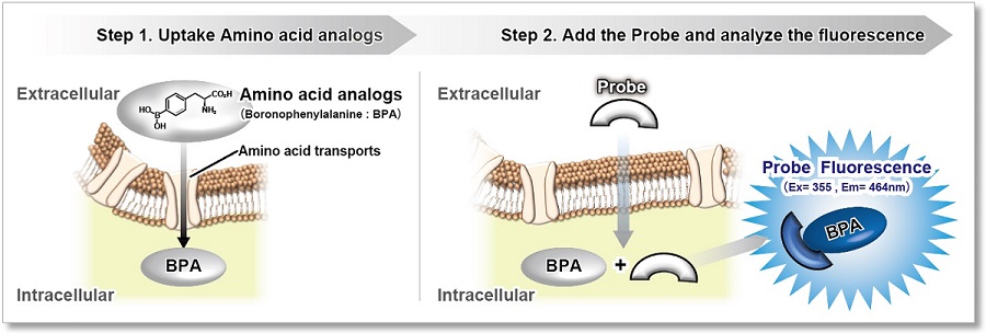
After amino acid analogs (BPA) are taken up into cells via amino acid transporters, the fluorescent probe permeates the cell membrane and binds to the amino acid analogs, emitting fluorescence (λex=360 nm, λem=460 nm). This product is suitable for fluorescence imaging, plate reader, and flow cytometry, making it possible to visualize and quantify the amino acid uptake capacity of cells and is useful for evaluation of amino acids uptake capacity and screening of amino acids transporter inhibitors.

Manual
Technical info
Evaluate the inhibitor of amino acids transporter and compare the amino acids uptake capacity between different cell lines. 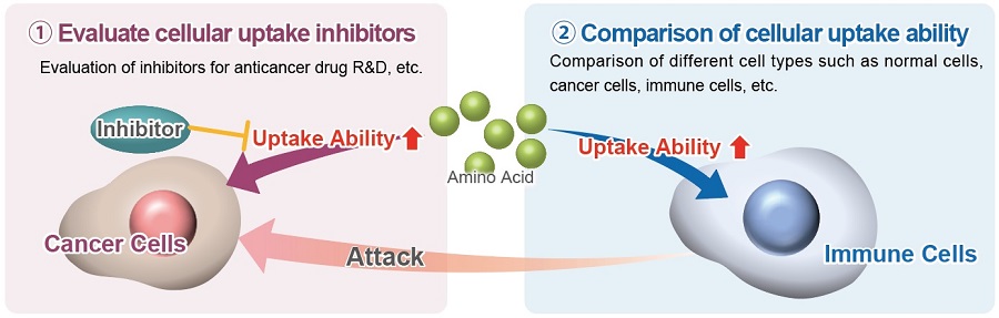
Comparison with Existing Method
Compare to the existing method, the required time can be greatly reduced.
| Method | Instrument | Time | Remarks |
|---|---|---|---|
|
Amino Acid Uptake Assay Kit |
Fluorescent Microscope |
60 min |
Simple procedure for amino acid analogs uptaking analysis |
|
Radioisotope Labeling |
Liquid Scintillation | 2 Days |
Allow testing different types of amino acids, |
| Metabolomics | LC/MS | 3 hr |
Allow testing different types of amino acids at the same time, |
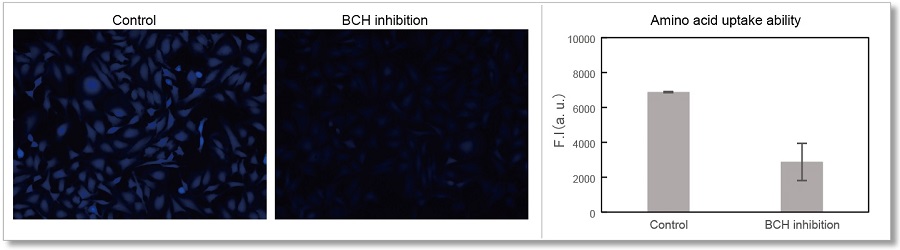
Application Data: Evaluation of BCH (Inhibitor of amino acids transporter)
Evaluate the inhibition ability BCH (2-Aminobicyclo[2.2.1]heptane-2-carboxylic acid) in HeLa cells with Amino Acid Uptake Assay Kit.
<Experimental Conditions>
Cell Line: HeLa cells
Medium: MEM (5.5 mmol/l Glucose)
Incubation: 1 mmol/l BCH/HBSS (Hanks' Balanced Salt Solution), 37℃, 30 min
Instrument:Fluorescent Microscopy(Ex=340-380 nm, Em: 435-485 nm)
Instrument:Plate Reader (Ex=360 nm, Em: 460 nm)

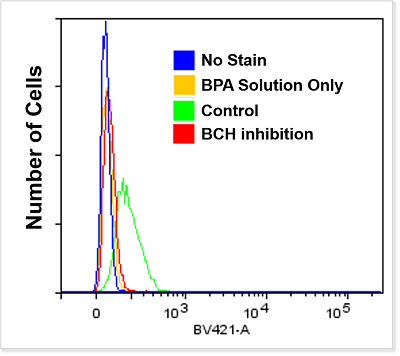
<Experimental Conditions>
Instrument: Flow Cytometer (Ex=405 nm, Em: 425-475 nm)
Branched Chain Amino Acids (BCAA) and Disease
It is clear that the intracellular metabolism of branched-chain amino acids (BCAA) is involved in various diseases such as cancer, aging, diabetes, and obesity. BCAA is mainly taken up into cells via neutral amino acid transporter (LAT).
The amino acid analog (BPA) used in this kit has been confirmed to be taken up via LAT in the same way as BCAA, and this kit can be used to evaluate the intracellular uptake of BCAA.
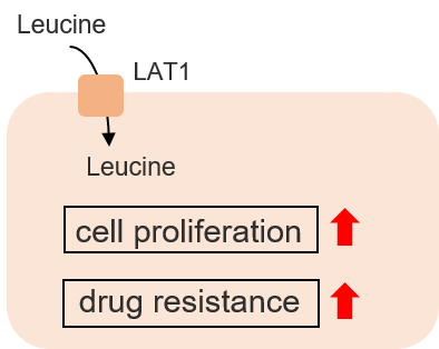
Cancer cells
Recent studies have shown that breast cancer cells take up large amounts of leucine, a BCAA, via LAT1. the BCAA uptake transporter LAT1 is expected to be a target for new breast cancer drugs.
Saito, Y., Li, L., Coyaud, E., et al, "LLGL2 rescues nutrient stress by promoting leucine uptake in ER+ breast cancer." Nature, 2019, 569, 275.
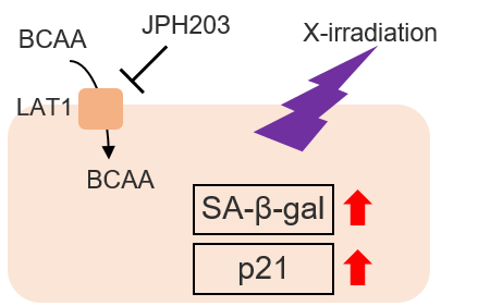
Cellular senescence
Cells are known to undergo cellular senescence upon X-irradiation via DNA damage and ROS generation. The combination of X-irradiation and LAT1 inhibitors in cancer cells has been shown to promote cellular senescence. The importance of BCAAs in cellular senescence has been suggested.
Tomoki Bo et al.,” LAT1 inhibitor JPH203 sensitizes cancer cells to radiation by enhancing radiation-induced cellular senescence”, Translational Oncology, 2021, 14, 101212.
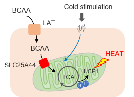
Obesity
Brown adipose tissue (BAT) during cold exposure takes up BCAAs for heat production. It has been shown that intracellularly taken-up BCAAs are oxidized in mitochondria and contribute to the heat production of BAT.
Yoneshiro T, et al., “BCAA catabolism in brown fat controls energy homeostasis through SLC25A44”, Nature, 2019, 572, 614.
Experimental Example: Evaluation with BCAA (Leucine)
The addition of L-Leucine, a BCAA, and BCH, a LAT1 inhibitor, inhibited the uptake of BPA into the cells.
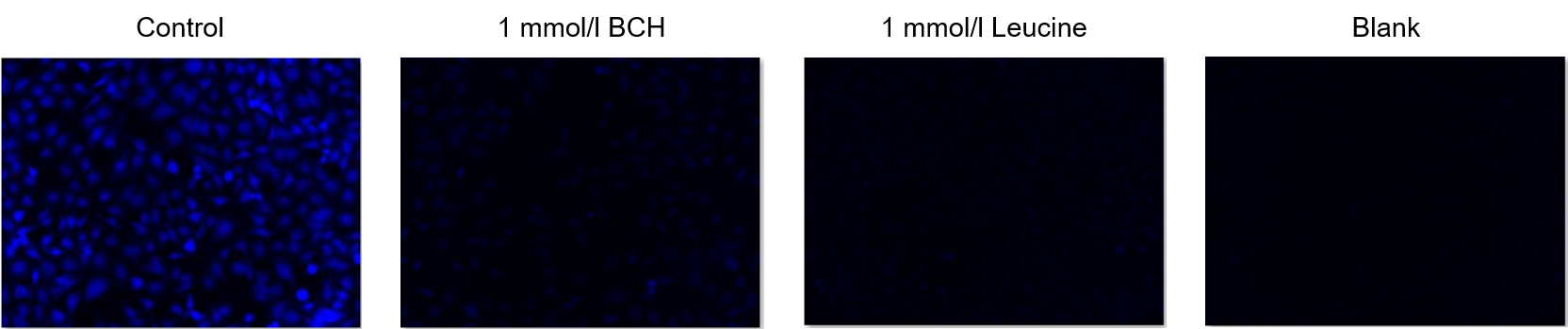
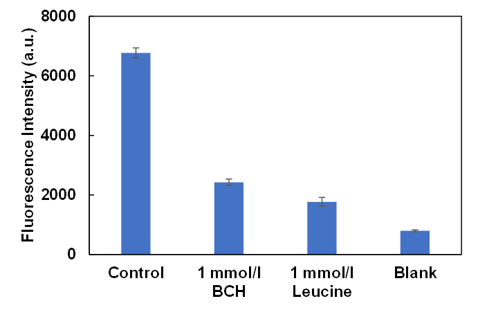
Cells: HeLa
Inhibitors: LAT1 inhibitor BCH (2-Aminobicyclo[2.2.1]heptane-2-carboxylic acid) and L-Leucine
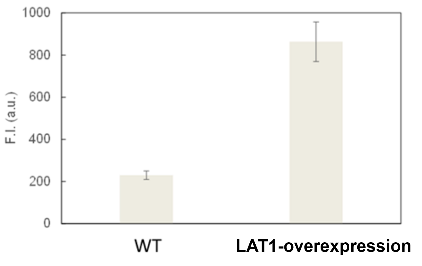
Experimental Example: Evaluation of Amino Acid Transporter Overexpressing Cells
Amino acid uptake in LAT-1-overexpressing MCF7 cells was greatly increased compared with the wild type.
Courtesy of Dr. Yasuhiro Saito (Institute for Advanced Biosciences, Keio University)
Experimental Example: Evaluation by Amino Acid Transporter Inhibitor (T3)
BPA cellular uptake was inhibited in a concentration-dependent manner by the addition of T3, an LAT1 inhibitor.
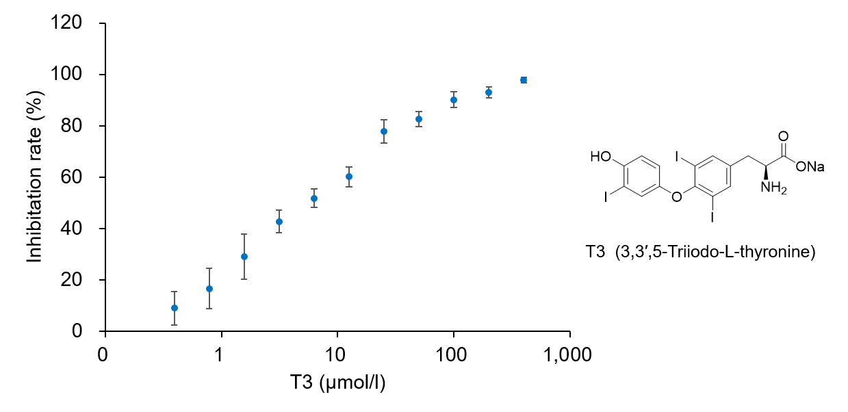
Cells: HeLa
Inhibitors: LAT1 inhibitor T3 (3,3′,5-Triiodo-L-thyronine)
Experimental Examole: Induction of Ferroptosis by Erastin
Erastin is a known inducer of ferroptosis. By inhibiting the cystine transporter (xCT), erastin inhibits the uptake of cystine. Cystine is the raw material for GSH. Therefore, Erastin ultimately decreases the amount of GSH. Decreased GSH then results in lipid peroxide accumulation and induction of ferroptosis.
The following experimental examples show changes in each aforementioned index as a consequence of erastin stimulation. Measurements are made using Dojindo reagents.
Using erastin-treated A549 cells, we measured intracellular Fe2+, ROS, lipid peroxide, glutathione, glutamate release into the extracellular space, and cystine uptake. As a result, inhibition of xCT by elastin was observed and also the release of glutamate and uptake of cystine were decreased. Furthermore, elastin treatment decreased intracellular glutathione while it increased intracellular Fe2+ , ROS, and lipid peroxides.


| 1 Amino Acid Uptake | : Amino Acid Uptake Assay Kit (Code: UP04) |
| 2 Glucose Uptake | : Glucose Uptake Assay Kit-Green (Code: UP02) |
| 3 Cystine Uptake | : Cystine Uptake Assay Kit (Code: UP05) |
| 4 Intracellular glutathione | : GSSG/GSH Quantification Kit (Code: G257) |
| 5 Intracellular labile Fe | : FerroOrange (Code: F374) |
| 6 Intracellular total ROS | : ROS Assay Kit -Highly Sensitive DCFH-DA- (Code: R252) |
| 7 Lipid Peroxides | : Liperfluo (Code: L248) |
Cell Line: A549
Incubation Conditions: 100 μmol/l Erastin/MEM, 37℃, 3h
References
| No. | Sample | Instrument | Reference |
|---|---|---|---|
| 1 | SCC7 Cell | Flow Cytometer |
T. Watanabe, Y. Sanada, Y. Hattori, and M. Suzuki, "Correlation between the expression of LAT1 in cancer cells and the potential efficacy of boron neutron capture therapy", 2022, J. Radiat. Res., doi:10.1093/jrr/rrac077. |
| 2 | CD4+ Cells | Flow Cytometer |
W. Zhang, X. Cao, X. Zhong, H. Wu, Y. Shi, M. Feng, Y.C. Wang, D. Ann, Y. Gwack, Y.C. Yuan, W. Shang and Z. Sun , "SRC2 controls CD4+ T cell activation via stimulating c-Myc-mediated upregulation of amino acid transporter Slc7a5", 2023, PNAS, doi:10.1073/pnas.2221352120. |
Q & A
-
Q
What microplates do you recommend?
-
A
We recommend the following microplates.
Company Product Name Cat No. ibidi μPlate 96 well ibiTreat black S 15 lb89626 AGC techno glass EZVIEW Glass Bottom Culture Plate LB 96well 5866-096 Thermo Fisher 96 Well Black/Clear Bottom Plate, TC Surface, Pack of 10 165305 How the type of plate affects the results, please refer to [Q&A: Does the type of microplate affect the results?] for more information.
-
Q
Does the type of microplate affect the results?
-
A
Yes. Not all microplates are compatible with this assay, and some might not enable certain measurements to be made (see reference data).
The following plates are recommended for use of this kit.Company Product Name Cat No. ibidi μPlate 96 well ibiTreat black S 15 lb89626 AGC techno glass EZVIEW Glass Bottom Culture Plate LB 96well 5866-096 Thermo Fisher 96 Well Black/Clear Bottom Plate, TC Surface, Pack of 10 165305
<Reference: Comparison between microplates>
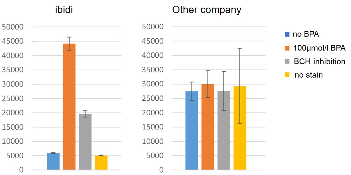
Using Ibidi plates and plates from other manufacturers, we investigated the ability of HeLa cells to uptake amino acids in the presence of BCH (2-Aminobicyclo[2.2.1]heptane-2-carboxylic acid), an inhibitor of amino acid transporters. However, we could not confirm the inhibition of uptake by BCH due to the higher background observed in the plates from other manufacturers compared to the recommended Ibidi plates.Fluorescence microscope
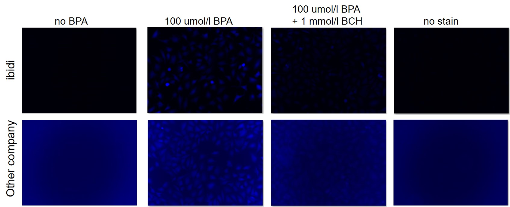
Plate reader
-
Q
Which transporter take BPA into cells?
-
A
BPA has been reported to be uptaken via LAT1, LAT2 and ATB0,+
(Reference: Wongthai P et al., “Boronophenylalanine, a boron delivery agent for boron neutron capture therapy, is transported by ATB0,+, LAT1 and LAT2”, Cancer Sci., 2015, Mar;106(3):279-86 )In addition, Dojindo has conducted a competitive inhibition assay with BCH, an inhibitor of LAT1, and leucine, a substrate of LAT1. The results indicated that BPA uptake is inhibited.
-
Q
Which cell lines has been analysis by this kit?
-
A
Adherent Cells include Hela, A549, HepG2, MCF-7, C2C12, MEF, U251.
Floating Cels include MOLT4.
-
Q
Where is BPA localized in Cells?
-
A
BPA is uniformly present in the cells after permeating the cell membrane.
-
Q
Is it possible to store the BPA solution and working solution for a period of time?
-
A
BPA solution and working solution cannot be stored.
-
Q
What should I do if there is no difference in the fluorescence signal?
-
A
There are two possible causes, which may be improved by the following.
(1) The uptake capacity of the BPA uptake solution may be low. In this case, please consider increasing the concentration of the BPA Solution. (5 to 50 times dilution).
(2) The working solution may have expired. Please prepare the working solution again.
-
Q
What should I do if the background is high?
-
A
There might be BPA remaining in the culture. In this case, please wash the cell once with HBSS.
-
Q
Is it possible to quantitative evaluation with BPA?
-
A
It is not possible to quantitative the probe uptaken by cells. This product is used to evaluate the increase or decrease of amino acid uptake ability.
-
Q
After BPA enters cells, is it broken down and metabolize
-
A
BPA has a stable structure and is not degraded after uptake.
-
Q
Is it possible to fix cells after incorporating BPA into living cells?
-
A
Cells cannot be fixed after staining because the probe leaks out of the cells.
Handling and storage condition
Related products
The following related products are also used in research related to this product.
-
Glucose uptake capacity assay
Glucose Uptake Assay Kit-Green
-
Glucose Metabolism Assay
Glucose Assay Kit-WST
-
Lactic Acid Measurement
Lactate Assay Kit-WST
-
α-Ketoglutaric Acid Measurement
α-Ketoglutarate Assay Kit-Fluorometric
-

Senescence Cell Detection (for Microplate)
Cellular Senescence Plate Assay Kit - SPiDER-βGal


