| This article focusing on the role of cholesterol accumulation in lysosomes of senescent cells, and its connection to maintaining the senescence-associated secretory phenotype (SASP). We believe that these findings can provide valuable insights into the relationship between senescence and lysosomal function. | |
|
Lysosomal control of senescence and inflammation through cholesterol partitioning Point of Interest |
|
| Related Techniques | |
| Cellular senescence detection (Live cell imaging or FCM) | Cellular Senescence Detection Kit HOT |
| Cellular senescence detection (Plate reader) | Cellular Senescence Plate Assay Kit HOT |
| Autophagy detection | DAPGreen / DAPRed (Autophagosome detection), DALGreen (Autolysosome detection) |
| Lysosomal function assay | Lysosomal pH and mass detection Kit |
| Lysosome staining | pH-dependent (Red) and pH-independent (Green / Deep Red) probes |
| Nutrient uptake Assay | Glucose (Blue / Green / Red), Amino Acid, Cystine, and Fatty Acid Uptake Assay Kit |
| Lipid droplets detection | Lipi-Blue / Green / Red / Deep Red |
| Related Applications | |
| Analysis of Lysosomal Mass and pH Exchange in Senescence-induced Cells | |
|
|
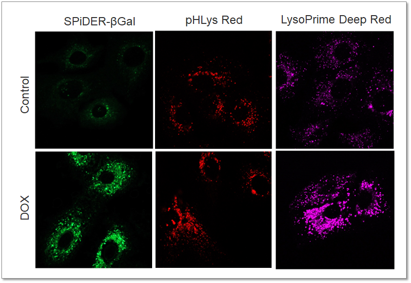
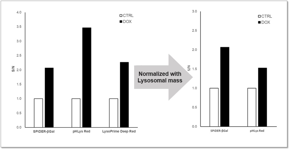
Purpose: To investigate changes in lysosomal mass and pH in A549 cells induced to senescence by treatment with Doxorubicin (DOX). Methods: Senescence-associated acidic β-galactosidase (SA-βGal) activity was detected using Cellular Senescence Detection Kit - SPiDER-βGal. Lysosomal mass was detected using LysoPrime Deep Red, and pH was detected using pHLys Red. Fluorescence imaging was used to observe changes in lysosomal mass and pH in senescent cells compared to non-senescent cells. The normalized fluorescence intensity of lysosomal mass and pH was also measured by a plate reader. Results: Our findings indicate that senescence induced by DOX resulted in an increase in lysosomal mass and acidification of pH compared to non-senescent cells. The obtained results are consistent with previous reports* that demonstrated enhanced lysosomal activity in senescent cells induced by the CDK4/6 inhibitor, palbociclib. The fluorescence imaging and plate reader data both support these findings. * Miguel Rovira, et. al., Aging Cell (2022)
<Experimental Conditions for Microscopy> <Experimental Conditions for Plate Reader>
<Products in Use> |