|
Autophagy plays a critical role in neurodegeneration and aging by maintaining cellular health through the degradation and recycling of damaged cellular components. In the context of neurodegeneration, such as Alzheimer's, Parkinson's and Huntington's diseases, impaired autophagy contributes to the accumulation of toxic protein aggregates that exacerbate neuronal damage and dysfunction. Conversely, enhancing autophagy has been shown to mitigate these deleterious effects and improve neuronal survival, suggesting a protective role in aging and neurodegenerative diseases. Research continues to explore autophagy modulators as potential therapeutic agents to slow the progression of neurodegenerative diseases and promote healthier aging.
|
-
The aging mouse CNS is protected by an autophagy-dependent microglia population promoted by IL-34
Click here for the original article: Rasmus Berglund, et. al., Nature Communications, 2024.
Point of Interest
- A microglial population emerges in the cortical regions of aging mice, characterized by a transcriptome indicative of activated autophagy.
- This population is found to be dependent on IL-34, a ligand for CSF1R.
- Loss of autophagy-dependent microglia leads to neuronal and glial cell death and increased mortality in aged mice exposed to neuroinflammation.
- Conversely, IL-34-mediated microglial expansion is protective.
-
Mitophagy curtails cytosolic mtDNA-dependent activation of cGAS/STING inflammation during aging
Click here for the original article: Juan Ignacio Jiménez-Loygorri, et. al., Nature Communications, 2024.
Point of Interest
- Mitophagy is either increased or unchanged in older mice compared to younger ones.
- There is a marked upregulation of the type I interferon response in the retinas of older mice.
- This response correlates with increased levels of cytosolic mtDNA and activation of the cGAS/STING pathway.
- In older mice, induction of mitophagy with urolithin A attenuates cGAS/STING activation and ameliorates the deterioration of neurological function.
-
SKA2 regulated hyperactive secretory autophagy drives neuroinflammation-induced neurodegeneration
Click here for the original article: Jakob Hartmann, et. al., Nature Communications, 2024.
Point of Interest
- SKA2 inhibits SA-dependent IL-1β release by counteracting FKBP5 function.
- Hippocampal Ska2 knockdown in male mice hyperactivates secretory autophagy (SA), leading to neuroinflammation and subsequent neurodegeneration.
- Hyperactivation of SA increases IL-1β release, contributing to an inflammatory feed-forward vicious cycle including NLRP3 inflammasome activation and Gasdermin D-mediated neurotoxicity.
- Results from protein analyses of postmortem human brains show that SA is hyperactivated in Alzheimer's disease.
|
|
Related Techniques
|
- First-time autophagy research
- Autophagic Flux Assay Kit
|
- Autophagy detection dyes for imaging
- DAPRed (Autophagosome detection), DALGreen (Autolysosome detection)
|
- Autophagy detection for Flow cytometry / plate assay
- DAPGreen (Autophagosome detection)
|
- Mitophagy detection dye
- Mitophagy Detection Kit and Mtphagy Dye
|
- Lysosomal pH and mass detection
- Lysosomal Acidic pH Detection Kit-Green/Red and Green/Deep Red
|
- Cellular senescence detection
- SPiDER-βGal for live-cell imaging or flow cytometry / microplate reader / tissue samples.
|
- Glycolysis/Oxidative phosphorylation Assay
- Glycolysis/OXPHOS Assay Kit
|
|
Related Applications
|
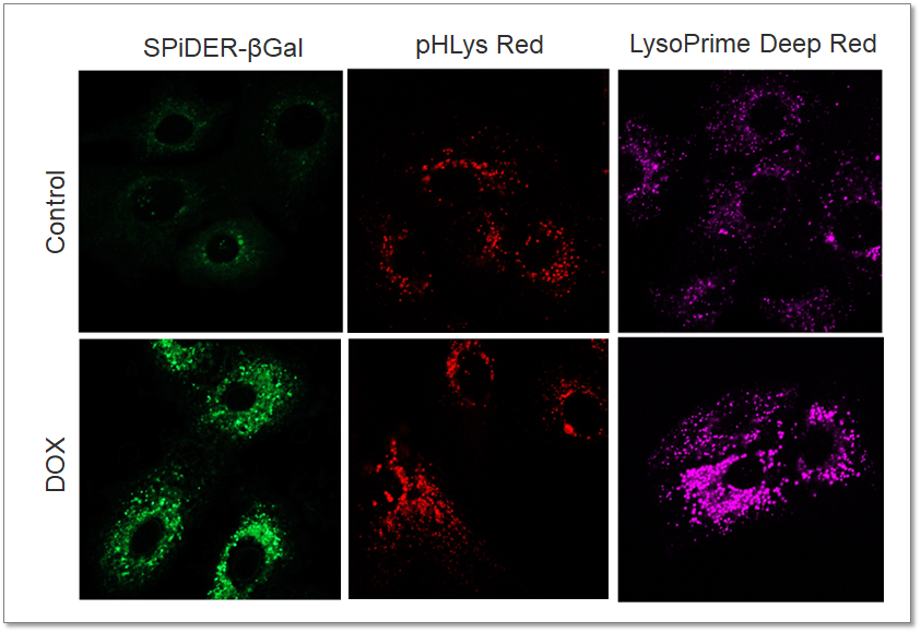
Analysis of Lysosomal Mass and pH Exchange in Senescence-induced Cells
-
Purpose: To investigate changes in lysosomal mass and pH in A549 cells induced to senescence by treatment with Doxorubicin (DOX).
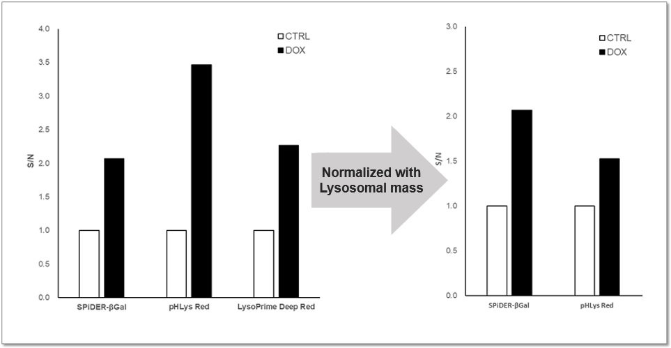
Methods: Senescence-associated acidic β-galactosidase (SA-βGal) activity was detected using Cellular Senescence Detection Kit - SPiDER-βGal. Lysosomal mass was detected using LysoPrime Deep Red, and pH was detected using pHLys Red. Fluorescence imaging was used to observe changes in lysosomal mass and pH in senescent cells compared to non-senescent cells. The normalized fluorescence intensity of lysosomal mass and pH was also measured by a plate reader.
Results: Our findings indicate that senescence induced by DOX resulted in an increase in lysosomal mass and acidification of pH compared to non-senescent cells. The obtained results are consistent with previous reports* that demonstrated enhanced lysosomal activity in senescent cells induced by the CDK4/6 inhibitor, palbociclib. The fluorescence imaging and plate reader data both support these findings.
<Experimental Conditions for Microscopy>
SA-βGal(Green):Ex = 488 nm, Em = 490 – 550 nm
Lysosomal pH (Red):Ex = 561 nm, Em = 560 – 620 nm
Lysosomal mass (Deep Red):Ex = 633 nm, Em = 640 – 700 nm
<Experimental Conditions for Plate Reader>
SA-βGal: Ex = 525 – 535 nm, Em = 550 – 570 nm
Lysosomal pH: Ex = 555 – 565 nm, Em = 590 – 610 nm
Lysosomal mass: Ex = 645 – 655 nm, Em = 690 – 710 nm
<Products in Use>
- Cellular Senescence Detection Kit
- Lysosomal pH and mass detection Kit
> More about Lysosomal Function Analysis
|


