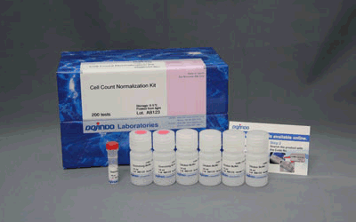
Cell Count Normalization Kit
Cell Count Normalization
- Enhance reliability of data on plate assay
- No need for lysis procedure
- Easily determine cell number just by adding the reagent
-
Product codeC544 Cell Count Normalization Kit
| Unit size | Price | FUJIFILM Wako Products code |
|---|---|---|
| 200 tests | ¥9,000 | 342-09393 |
| 1000 tests | ¥22,000 | 346-09391 |
| 200 tests | ・Staining Solution ・Dilution Buffer ・Quenching Buffer |
50 μl×1 10 ml×4 10 ml×2 |
|---|---|---|
| 1000 tests | ・Staining Solution ・Dilution Buffer ・Quenching Buffer |
250 μl×1 100 ml×2 100 ml×1 |
Product Description
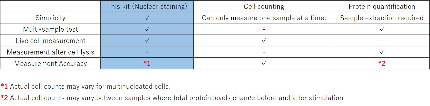
Cell Count Normalization Kit employs a nucleic acid staining dye, Hoechst 33342 which binds to nuclear DNA to emit blue fluorescence. By measuring this blue fluorescence, correction of the measured value can easily be carried out in simple steps whereas the visual cell counting method requires cumbersome procedure. Moreover, unlike the correction by protein or ATP amount, the kit requires no lysis procedure. In addition, Quenching Buffer included in the kit enables a direct measuring of fluorescence signal without any background.
Technical info
When cells are analyzed in a microplate, the results obtained may sometimes differ depending on the number of cells per well. In such cases, normalization of the measured values will be necessary.
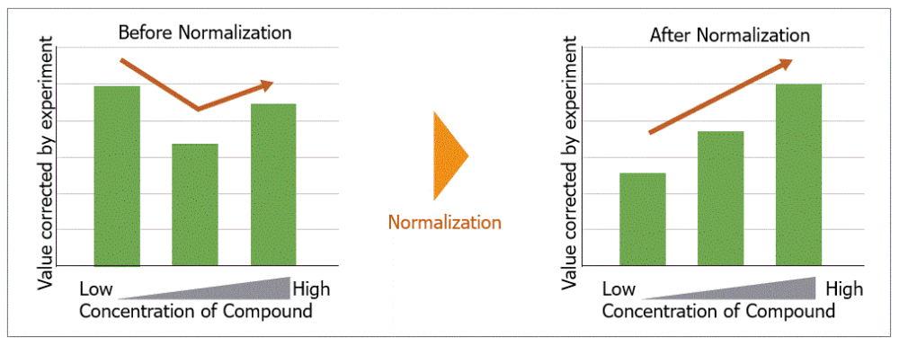
Comparison with conventional method
Normalization of the measured values at the time of plate assay is done by using the cell number or nucleus/protein level as an indicator. Among these detection techniques, our nuclear staining kit is easy to use and capable of analyzing multiple specimens.
High correlation with cell number
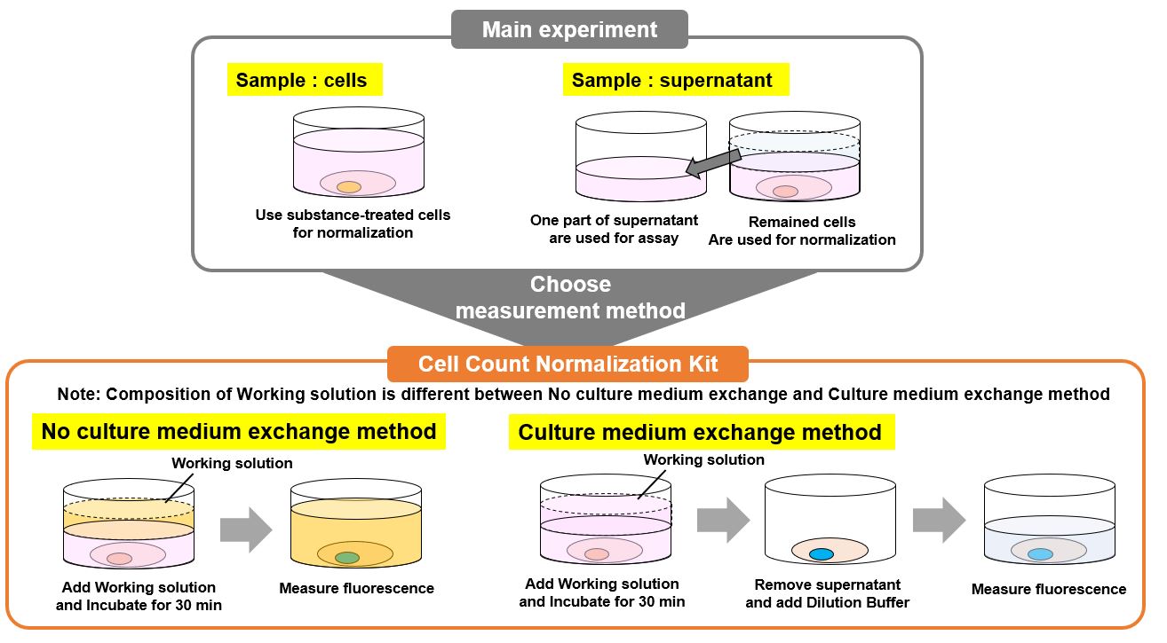
When you are performing a plate assay the data needs to be normalized. In order to normalize the data, an assessment method equivalent (i.e. cell counting) is essential. In the experiments below, changes in fluorescence intensity were confirmed by cell number-dependents. Data at the time of plate assay were normalized by using this kit and the cell counting method. A comparison was then made between the two methods.
Cell number-dependent changes in fluorescence intensity
Cell number-dependent changes in fluorescence intensity were observed, and the results were highly linear.
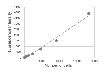
HeLa cells were serially diluted and seeded in a 96-well microplate. After incubation overnight, fluorescence intensity was measured using this kit.
Comparison with cell counting method
When normalization occurred at the time of the plate assay (lactate measurement), this kit showed equivalent results to cell counting.
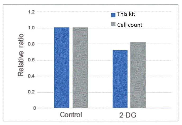
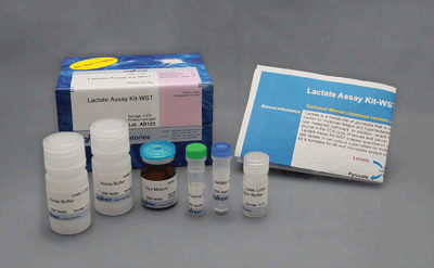
2-Deoxy-D-glucose was added to HeLa cells. Lactate levels in the supernatant were quantified using the Lactate Assay Kit-WST (Item#: L256)
References
Q & A
-
Q
Is it possible to measure with other fluorescent dyes at the same time?
-
A
If the excitation and fluorescence wavelengths overlap with those of Hoechst 33342 used in this kit, evaluation by co-staining is not possible.
Please make sure that the excitation wavelength (300-400 nm) and fluorescence wavelength (400-550 nm) of Hoechst 33342 do not overlap.If the wavelengths do not overlap, use other fluorescence dyes for evaluation before using this kit.
Handling and storage condition
| 0-5°C, Protect from light |
Related products
The following related products are also used in research related to this product.