|
Extracellular vesicles mediate disease-related cell communication in cancer and neurodegeneration. Recent studies suggest that EV analysis should extend beyond total uptake to include target-cell entry routes, recipient-cell responses, and cargo degradation or release. Tumor-derived sEVs enter cells through defined, subtype-influenced routes and can trigger Ca²⁺ signaling that enhances uptake. In a neurodegeneration model, human microglia took up tau, an aggregation-prone protein linked to Alzheimer’s disease, but incompletely degraded fibrillar tau, releasing aggregation-inducing tau, partly in EV-associated form.
|
|
Uptake of small extracellular vesicles by recipient cells is facilitated by paracrine adhesion signaling
(Nature communications, 2025)
Summary:
This study directly observed how individual tumor-derived sEVs enter recipient cells using live-cell imaging. The results showed that sEV uptake is not simply a matter of total amount, but depends on the type of sEV, the route used to enter the cell, and signaling responses in the recipient cell. These findings suggest that cancer EV studies should evaluate not only whether EVs are internalized, but also how they enter cells and whether they activate recipient-cell signaling.
Highlighted technique:
To evaluate sEV uptake and signaling in recipient cells, the authors isolated sEVs from cultured donor-cell media, fluorescently labeled sEVs or sEV subtypes, and tracked their internalization by live-cell imaging. Intracellular Ca²⁺ responses after sEV binding were measured by fluorescence Ca²⁺ imaging.
For EV uptake and intracellular response analysis, ultracentrifugation-free exosome isolation kit, exosome membrane labeling kit, and intracellular Ca²⁺ ion measurement kit are available.
|
|
Tracking tau and cellular responses in human iPSC-microglia: from uptake to seedable secretion, including in extracellular vesicles
(Alzheimer's & Dementia, 2026)
Summary:
This study showed that human iPSC-derived microglia handle extracellular tau, an aggregation-prone protein linked to Alzheimer’s disease, differently depending on tau form. Unlike monomeric tau, fibrillar and Alzheimer’s brain-derived tau resisted degradation and altered microglial response states, while incompletely processed fibrillar tau was released in forms that could induce tau aggregation in neurons, including EV-associated tau. These findings suggest that neurodegeneration studies should assess tau uptake together with degradation capacity, microglial responses, and the activity of released tau.
Highlighted technique:
To assess microglial handling of extracellular tau, human iPSC-derived microglia-like cells were exposed to monomeric, fibrillar, or Alzheimer’s brain-derived tau. The study measured tau uptake, tau-induced transcriptional responses, LDH-based cytotoxicity after brain-derived tau exposure, and extracellular vesicle-associated tau release using imaging, flow cytometry, RNA-seq, and EV assays.
To support research on extracellular tau uptake, microglial responses, and EV-mediated release, tools are available for evaluating endocytosis-related uptake processes and LDH-based cytotoxicity after tau exposure.
|
Application Note
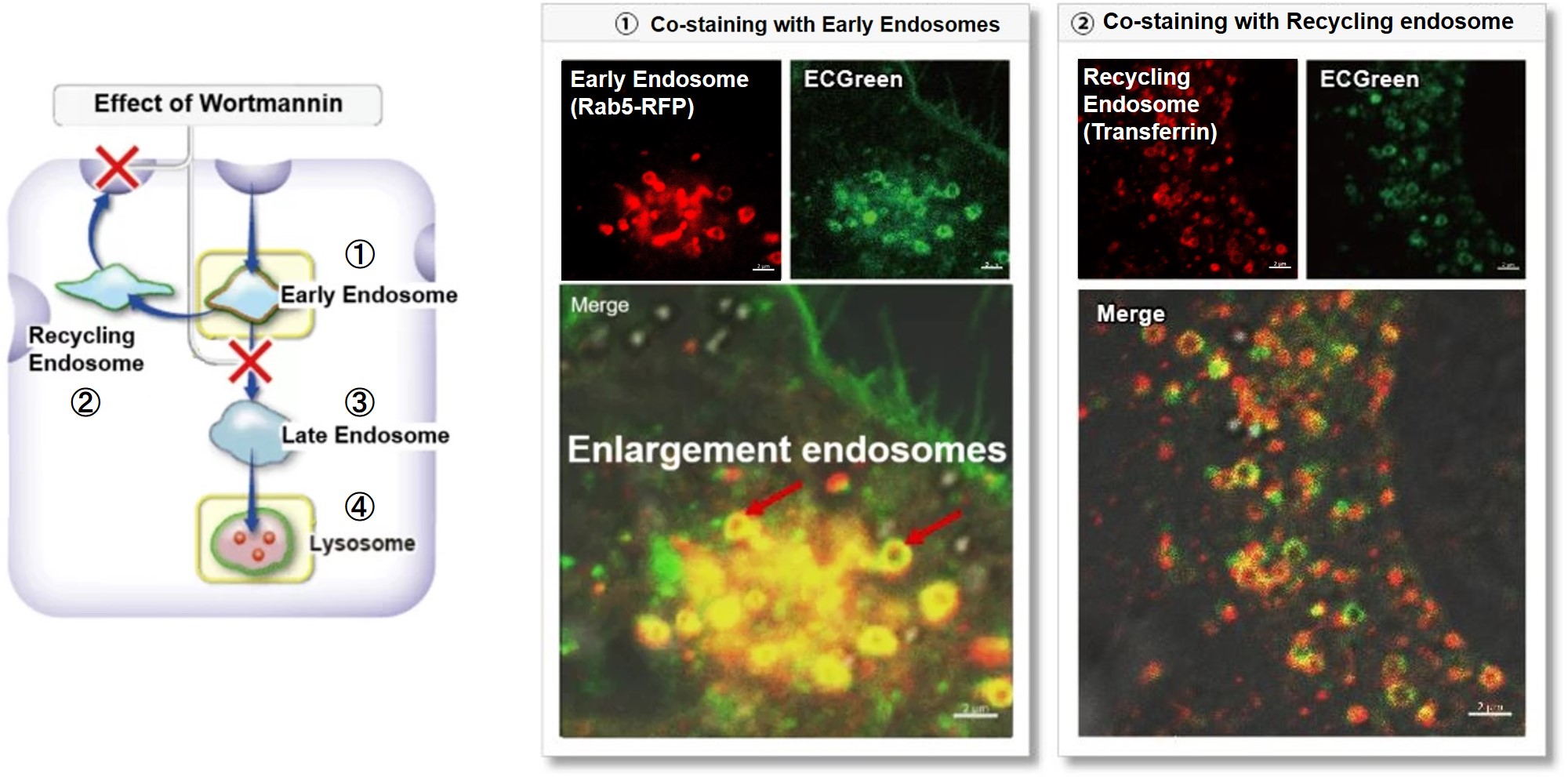
> Clear visualization of intracellular vesicular trafficking
Wortmannin is known to inhibit endosomal recycling and lysosomal translocation, leading to endosomal enlargement.
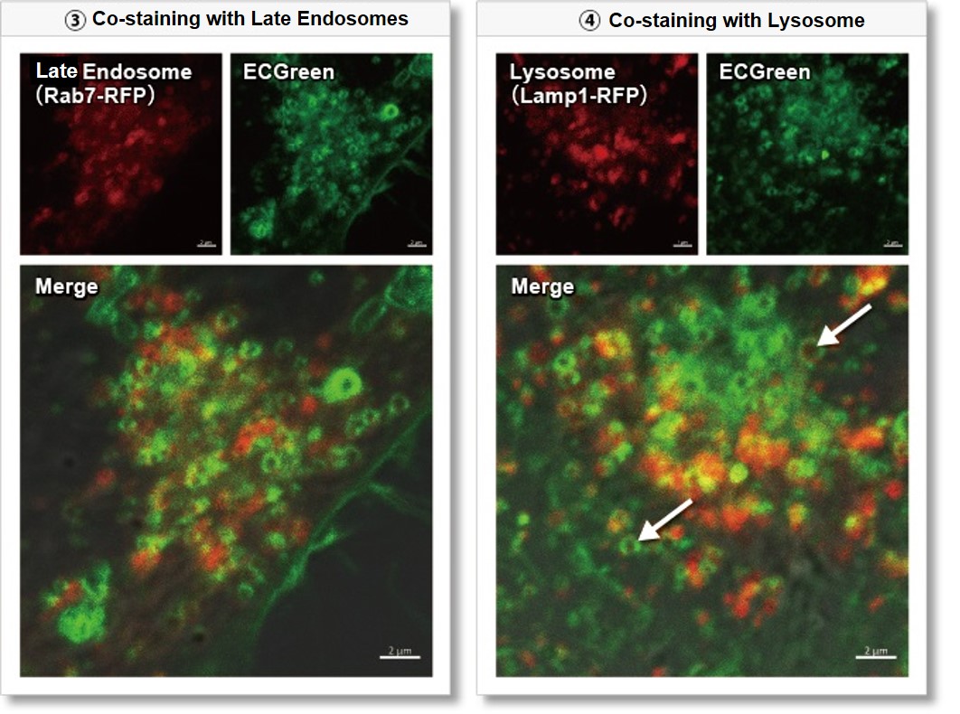
These changes induced by Wortmannin were confirmed by co-staining with ECGreen (green) and the following indicators.
①Eary endosome: Rab5-RFP (red)
② Recycling endosome: Fluorescent labeled Transferin (red)
③ Late endosome: Rab5-RFP (red)
④ Lysosome: Lamp1-RFP (red)
As a result, it was confirmed that ECGreen (green) co-localizes only with enlarged early endosomes and recycling endosomes (Fig. ① and ②), but not with late endosomes or Lysosomes (Fig. ③ and ④), supporting Wortmannin's effect. ECGreen can visualize changes in the intracellular vesicular trafficking system and endosome shape.
Endosomes (ECGreen, green): Ex. 405 nm / Em. 500 – 560 nm
Early endosomes (Rab5-RFP, red): Ex. 561 nm / Em. 560 – 620 nm
Recycling endosome (Transferrin-Alexa fluor 488 conjugate, red: pseudo-color): Ex. 488 nm / Em. 500 – 550 nm
Late endosomes (Rab7-RFP, red): Ex. 561 nm / Em. 560 – 620 nm
Lysosomes (Lamp1-RFP, red): Ex. 561 nm / Em. 560 – 620 nm
(1) Prepare HeLa cells in 8 wells of μ-Slide and incubate overnight.
(2) After washing with HBSS, 200 µl of Wortmannin (final concentration: 100 nmol/l) prepared in 10% FBS-containing MEM medium was added.
(3) Incubate at 37°C for 30 minutes
(4) 200 µl of ECGreen (diluted 1,000-fold) prepared in 10% FBS-containing MEM medium without removing the supernatant
(5) Incubate at 37°C for 30 minutes
(6) Wash the cells twice with HBSS and add MEM medium containing 10% FBS.
(7) Observation with a confocal laser microscope
|