| Neurons within and near tumors can interact with cancer cells and have been implicated in cancer progression. It is important to determine whether tumor-associated neurons directly supply metabolic support to cancer cells, as this has implications for elucidating mechanisms of metastasis and treatment response. Recent studies have investigated mitochondrial transfer from distinct donor cell types to cancer cells. One study demonstrated that tumor-associated neurons transfer mitochondria to cancer cells, sustaining bioenergetics, enhancing stress resistance, and promoting metastatic colonization. Another showed that adipose stem cell–derived mitochondria increase ATP production and ABC transporter–mediated drug efflux in breast cancer cells, reducing intracellular accumulation of chemotherapeutic agents. Together, these findings establish donor cell mitochondrial transfer as an experimentally quantifiable process that directly alters cancer cell behavior, including metastatic potential and drug resistance. |
|
Nerve-to-cancer transfer of mitochondria during cancer metastasis (Nature, 2025)
Summary:
This study investigated whether cancer-associated neurons can directly fuel cancer metabolism. Using coculture systems, mitochondrial transfer reporters, denervation models, and in vivo fate mapping, the authors demonstrated that neuron-derived mitochondria enhance cancer cell bioenergetics and stress resistance. Mitochondria-recipient cancer cells or their progeny were more frequently detected at metastatic sites, suggesting improved survival during metastatic stress.
Highlighted technique:
To evaluate whether neuron-derived mitochondria improve energetic and redox stress adaptation in cancer cells, the authors identified mitochondrial transfer using a newly developed genetic reporter and isolated the corresponding 4T1 cell populations by flow cytometry. Mitochondria-recipient cells showed increased total ATP, higher GSH, improved GSH/GSSG ratios, and reduced susceptibility to H2O2-induced cell death.
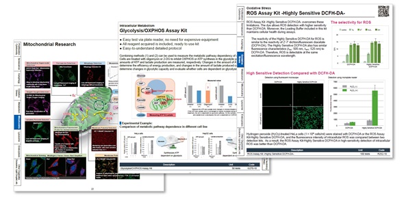
A mitochondrial staining reagent is available that enables continuous labeling for up to 7 days, making it useful for long-term tracking. Mitochondrial function and cellular redox status can be evaluated using parameters such as intracellular ATP, mitochondrial ROS and GSSG/GSH.
|
|
Mitochondrial transfer from Adipose stem cells to breast cancer cells drives multi-drug resistance (Journal of Experimental & Clinical Cancer Research, 2024)
Summary:
This study showed that adipose stem cells transfer mitochondria to breast cancer cells, thereby enhancing ATP production and upregulating ABC transporter-mediated drug efflux. As a result, breast cancer cells exhibited reduced intracellular drug accumulation, leading to increased resistance to chemotherapeutic agents such as doxorubicin, cisplatin, and docetaxel.
Highlighted technique:
To test whether ASC-derived mitochondria enhance ATP availability for ABC transporter-mediated drug efflux, isolated ASC-derived mitochondria were introduced into breast cancer cells. Seahorse OCR analysis measured mitochondrial respiration and ATP production, while intracellular doxorubicin accumulation was evaluated by its fluorescence intensity.
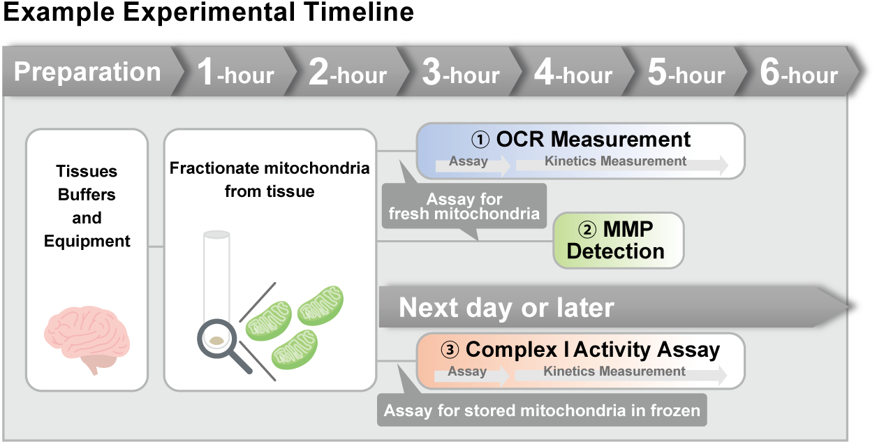
There is an OCR plate-assay kit that requires fewer cells and offers lower running costs. It can also be used for preliminary evaluation prior to Seahorse analysis. The mitochondrial isolation kit enables fractionation of intact mitochondria from tissue in approximately two hours.
|
Application Note I
> Inhibition of Mitochondrial Electron Transport Chain
|
|
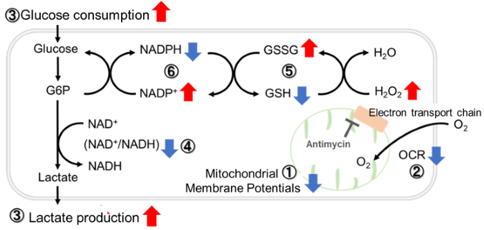
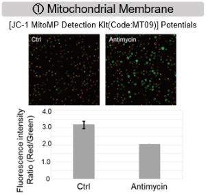
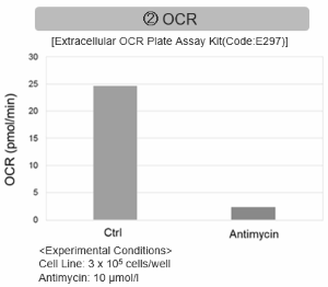
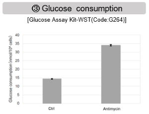
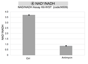
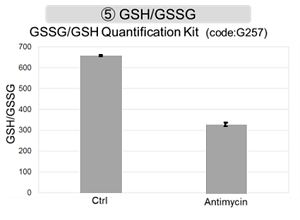
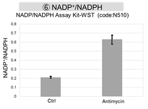
Antimycin stimulation of Jurkat cells was used to evaluate the changes in cellular state upon inhibition of the mitochondrial electron transport chain using a variety of indicators.
The results showed that inhibition of the electron transport chain resulted in (1) a decrease in mitochondrial membrane potential and (2) a decrease in OCR. In addition, (3) the NAD+/NADH ratio of the entire glycolytic pathway decreased due to increased metabolism of pyruvate to lactate to maintain the glycolytic pathway, (4) GSH depletion due to increased reactive oxygen species (ROS), and (6) increase in the NADP+/NADPH ratio due to decreased NADH required for glutathione biosynthesis were observed.
|
|
Application Note II
> Activity Evaluation of Mitochondria Fractionated from Mouse Brain
|
|
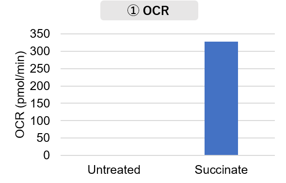
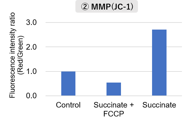
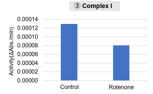
Mitochondria were isolated from mouse brain tissue, and oxygen consumption rate (OCR), mitochondrial membrane potential (MMP), and Complex I activity were measured.
The results showed that the addition of succinate, a substrate that activates Complex II of the electron transport chain, increased both OCR and MMP. In contrast, FCCP treatment reduced MMP, indicating that intact mitochondria were successfully fractionated.
Furthermore, in the Complex I activity assay, a decrease in activity was observed following treatment with rotenone, a Complex I inhibitor.
|
   |
<Product used>
Mitochondrial Fractionation: IntactMito Fractionation Kit for Tissue (Code: MT17)
OCR measurement: Extracellular OCR Plate Assay Kit (Code: E297)
MMP detection: JC-1 MitoMP Detection Kit (Code: MT09)
Complex I activity assay: MitoComplex- I Activity Assay Kit (Code: MT18) |
<Experimental Conditions>
OCR Measurement
Amount of mitochondria: 50 μg/well (as protein levels)
Succinate: 10 mmol/l
MMP Detection
Amount of mitochondria: 50 μg/well (as protein levels)
Succinate: 10 mmol/l, FCCP: 4 μmol/l
Complex I Activity Assay
Amount of mitochondria: 20 μg/well (as protein levels)
Rotenone: 10 μmol/l
|
|
| Mitochondrial and Metabolic Activity Indicators
|