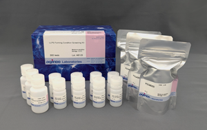
LLPS Starter Kit
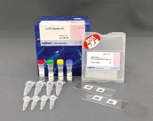
Ready-to-use Kit for The First Time LLPS Research
- All-in-one kit for making phase-separated droplets with BSA (Bovine Serum Albumin).
- Droplets are easy to observe using only a micropipette and microscope.
- Includes a user's manual with calculation examples and details on how to use it
-
Product codeLL01 LLPS Starter Kit
| Unit size | Price | Item Code |
|---|---|---|
| 1 set | $200 | LL01-10 |
| 1 set | BSA BSA Dissolving Buffer (20 mmol/l HEPES pH7.4, 150 mmol/l NaCl) Assay Buffer (60 mmol/l HEPES pH7.4, 450 mmol/l NaCl) 30% PEG8000 Solution Slide Glass with Double-Sided Tape Cover Glass 1.5 |
10 mg× 1 × 1 × 1 × 1 × 2 × 4 × 4 × 4 |
|---|
What is Liquid-Liquid Phase Separation (LLPS)?
Liquid-liquid phase separation (LLPS) is a phenomenon in which certain molecules assemble locally within a cell to form aggregates (droplets) of biomolecules with liquid-like properties. In recent years, LLPS has attracted much attention as it has been shown to affect many biological processes in the cell. Although the study of droplets formed by phase separation is still in its infancy, elucidating how these biological phenomena affect cellular functions and the pathogenesis of disease is considered key to the development of new therapeutic strategies.

Reference: E. Dolgin, Nature, 2018, DOI: 10.1038/d41586-018-03070-2.
Description
To understand phase separation in living organisms, researchers observe phase-separated droplets in cell-free systems using purified proteins. A common method involves labeling these proteins with fluorescent markers and observing their dynamics under a microscope. Additionally, researchers study the mechanisms of droplet formation by examining how droplets react in environments with different added chemicals. With many methods available, beginners might find it challenging to decide which approach to start with.
To help beginners, we have developed an all-in-one kit based on commonly used methods for droplet formation. This kit includes all necessary reagents and containers to allow basic microscopic observations of droplets formed by phase separation in vitro using BSA.
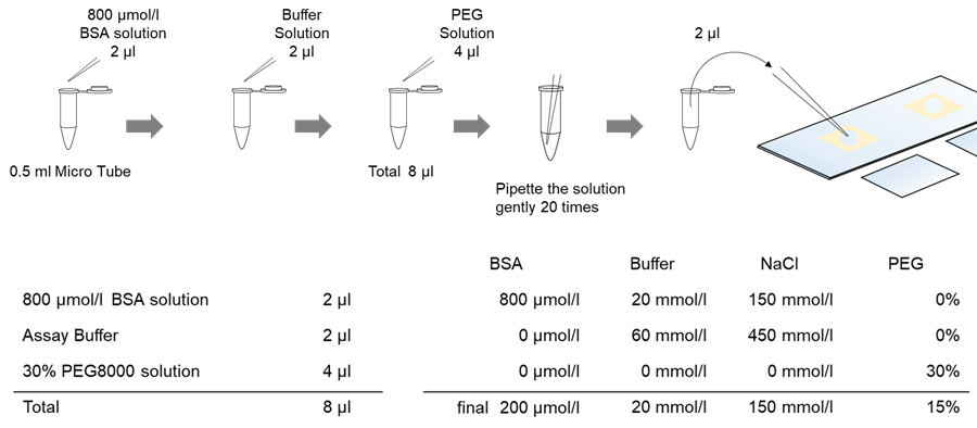
Figure 1. Procedure and composition table
Manual
Technical info
Generally, when starting LLPS research, it is often necessary to confirm that the target protein forms droplets in a cell-free system.
However, the optimal conditions for this step also differ depending on the protein, and there are various considerations and precautions such as pH, salt type, and the presence or absence of crowding agents*.
We have prepared two types of kits that are ideal for those who are just starting out in LLPS research.
*Crowding agent (molecular adulterant reproducer): A polymer such as PEG or Ficoll that is added to reproduce a crowded intracellular environment.
LL01 LLPS Starter Kit: Ready-to-use kit for the first time LLPS research
LL02 LLPS Forming Condition Screening Kit: Ideal for optimization of conditions for target protein droplets

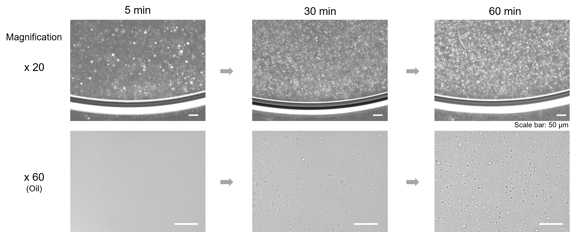
Observation of phase-separated droplets of BSA over time using this kit.
Phase-separated droplets of BSA were prepared according to the method described in the instruction manual, and changes over time were observed using a fluorescence microscope (model: BZ-X710: KEYENCE).
Observation of fusion of phase-separated droplets
The flowability of the BSA droplets was checked to confirm that they were phase-separated droplets.
Fusion of BSA phase separated droplets was observed under a microscope in bright field using the LLPS Starter Kit.
*Microscope used: KEYENCE BZ-X710 (60X magnification)

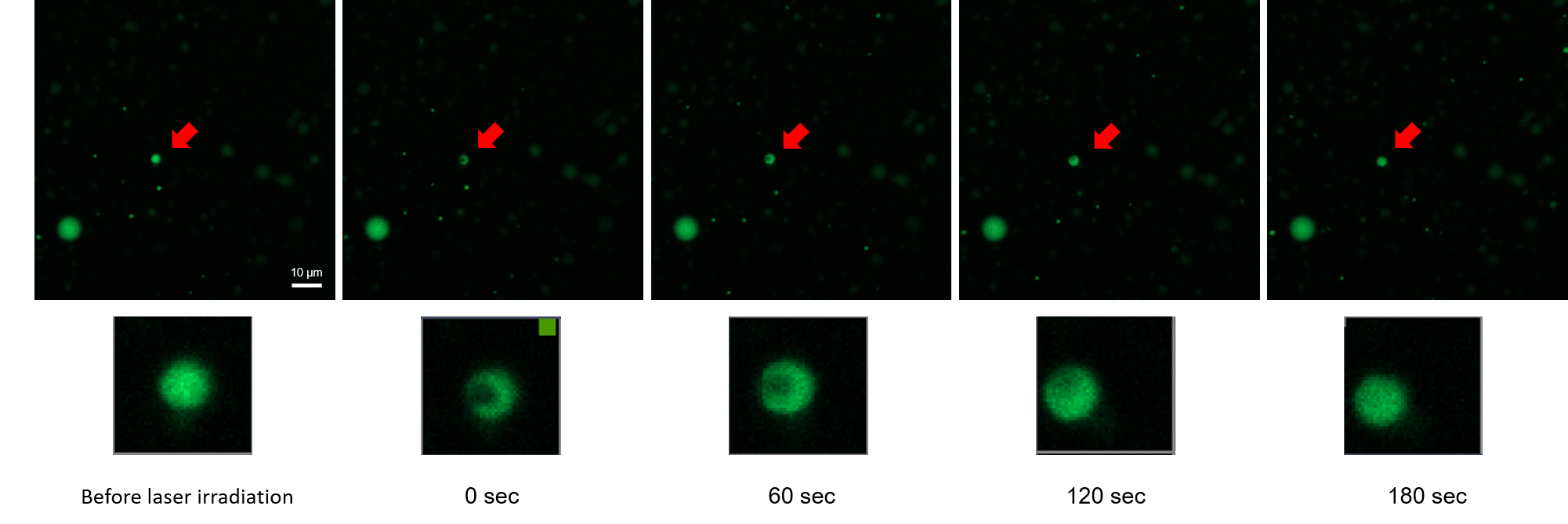
Fluorescence Recovery After Photodegradation (FRAP) measurements of phase-separated droplets
In the FRAP assay, a portion of the droplet is bleached by a laser and the fluorescence is observed to recover over time. If the bleached droplet is not liquid, the bleached portion remains intact. On the other hand, if the droplet is a liquid phase separated droplet, molecules from the surrounding area will enter the faded area and the fluorescence will recover. An example of FRAP measurement using this kit and our product (LK01) Fluorescein Labeling Kit - NH2 is shown below.
Phase-separated droplets were prepared using BSA labeled with a fluorescent dye, and FRAP measurement was performed 30 minutes later. After laser irradiation, the fluorescence of the bleached droplets was observed to recover.
*The final concentration of BSA in this experiment was 100 µmol/L. It was also prepared to contain 0.5% fluorescent dye labeled BSA.
For fluorescent dye labeling, 100 µg of BSA was labeled with (LK01) Fluorescein Labeling Kit - NH2 1 sample.
Microscope: Zeiss LSM 8 (63x magnification), Ex: 488 nm / Em: 500-600 nm, 0.5%, 500V
Q & A
-
Q
With one kit, how many observations can I make?
-
A
The glass slides provided in the kit can be used for 4 observations.
-
Q
Droplets cannot be observed.
-
A
Please check the following.
1. The number of phase-separated droplets may depend on the temperature. It is recommended that the experiment be conducted at room temperature of 20-25°C.
2. Before pipetting, if there is foaming or the solution adheres to the wall surface, it may not be mixed properly. Please use a tabletop centrifuge or similar device before pipetting. Also, pipetting should be done gently to avoid foaming.
3. Please extend the measurement time from 30 minutes, to 1 hour, to 2 hours after sample preparation.
4. Increase the concentration of BSA to 1000 µmol/l.
5. It is recommended to check the periphery of the solution first, as the edge of the drop of solution is easy to see the droplet. It is also recommended to increase the magnification of the microscope lens from 4x to 20x to 60x, checking the position of the phase-separated droplets.
-
Q
How can I confirm that it is a phase-separated droplet?
-
A
To prove that the observed object is a fluid droplet, observation of fusion of droplets, measurement of droplet sphericity, and FRAP measurement are used. For details, please refer to the Technical Information: Fluorescence Recovery After Photobleaching (FRAP) measurement of phase-separated droplets and observation of fusion of phase-separated droplets on this product page.


