Ab-10 Rapid Fluorescein Labeling Kit

Antibody Labeling
- Small Quantity of Antibody Requirement : 10 ug Antibody Sample Labeling
- Easy Labeling Procedure : Just Mix Antibody and Labeling Agent
- Rapid Labeling : Less than 30 minutes
-
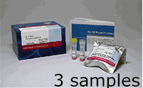
Product codeLK32 Ab-10 Rapid Fluorescein Labeling Kit
| Unit size | Price | Item Code |
|---|---|---|
| 3 samples | $330.00 | LK32-10 |
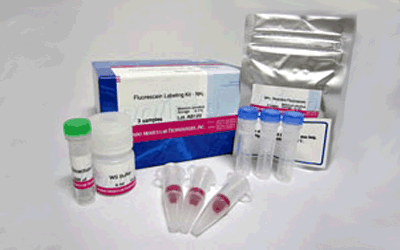
| 3 samples | ・Reactive Fluorescein ・Reaction Buffer ・Stop Solution |
x 3 100 μlx 1 100 μlx 1 |
|---|
Description
Ab-10 Rapid Fluorescein Labeling Kit is for labeling fluorescein to 10 µg antibody in less than 30 minutes. Labeling agent included in the kit has succinimidyl ester group , which can easily make a covalent bond with an amino group of the target antibody without any activation process. The kit contains all the necessary reagents for antibody labeling.

Fig. 1 IgG Labeling Reaction of NH2-reactive Fluorescein

Fig. 2 Labeing Procedure

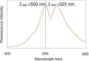
Fig. 3 Fluorescence Spectrum of Fluorescein
| Developer | Dojindo Molecular Technologies, Inc. |
|---|
Manual
Technical info
♦ Use 0.5-1 mg/ml of antibody solution for labeling. If the antibody concentration is higher than 1mg/ml, dilute the antibody solution with PBS.
♦ If the sample solution contains small insoluble materials, centrifuge the solution, and use the supernatant for the labeling.
♦ The microtube in this kit contain solutions. Since there is a possibility that the droplets might be attached to the inside walls or caps, please centrifuge to drop the droplets prior to opening.
♦ Some additives in antibody solution may interfere with the labeling if the concentration is too high. The maximum compatible concentrations of such additives are indicated on Table 1.
♦ After a Reactive Fluorescent is removed from the seal bag, keep the unused Reactive Fluorescein in the bag, seal tightly and store at -20ºC.Store the other components at 0-5ºC.
♦ Since reactive reagent binds to an amino group in antibody, there is a possibility that the labeled antibody loses the antigen recognition ability (Table 2).
Table. 1 Compatible concentrations of the additives

Table. 2 Non-Compatible Antibody

Experimental Data

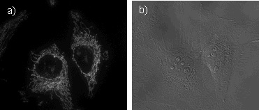
Fig. 3 Microscope Image of Mitochondria in Hela Cells a) Fluorescence Image b) Bright Field Image Fluorescence were to anti-mitochondria antibody using Ab-10 Rapid Fluorescein Labeling Kit (code:LK32)
References
1) K. Miyazaki, J. Oyanagi, D. Hoshino, S. Togo, H. Kumagai and Y. Miyagi, Cancer cell migration on elongate protrusions of fibroblasts in collagen matrix.', Sci Rep., 2019, 9, (1), 292.
Handling and storage condition
| 0-5°C, Protect from moisture |


