 Hidden sections will not be printed.
General Information
Reactive oxygen species (ROS) are mainly produced in mitochondria during ATP synthesis. ROS play an essential role in signaling pathways and the immune system, whereas increasing ROS is associated with diseases and cellular senescence. Recent studies suggested that ferroptosis is a new type of cell death characterized by iron dependency and increasing ROS; therefore ROS detection has been attracting considerable interests in ferroptosis research1).
DCFH-DA is widely used for ROS detection, but it has some limitations such as weak fluorescence signals and high background. Dojindo’s ROS Assay Kit -Highly Sensitive DCFH-DA- overcomes these limitations. The dye which is employed in the kit allows ROS detection with higher sensitivity than DCFH-DA. Moreover, the Loading Buffer included in this kit maintains cellular health during assays.
Kit Contents
| Highly Sensitive DCFH-DA Dye Loading Buffer (10×) |
10 μl ×1 1 ml ×1 |
Storage Condition
Store at -20℃
Required Equipment and Materials
- Fluorescence microscope, fluorescence plate reader, or flow cytometer
- Incubator (37℃)
- Micropipettes (100–1000 μl, 0.5–10 μl)
- Microtubes
- Hanks' Balanced Salt Solution (HBSS)
Precaution
Allow the ROS Assay Kit -Highly Sensitive DCFH-DA- to equilibrate at room temperature before use.
Centrifuge the tube briefly before opening the cap because the content may be on the tube wall or the ceiling of the cap.
Please refer to Table 1 to check suitable fluorescence wavelengths for each application.
Table 1. Recommended filter settings of the kit
| Applications | Fluorescence plate reader | Fluorescence microscope | Flow cytometer |
| Measurement wavelength | Excitation: 490–520 nm Emission: 510–540 nm |
・Confocal microscope Ex/Em: 488/500–550 nm ・Fluorescence microscope GFP or FITC filter |
FITC filter |
Preparation of Solutions
Preparation of Loading Buffer solution
Dilute the 10× Loading Buffer (1:10) with double-deionized water.
- Refer to Table 2 for preparing the Loading buffer solution.
Table 2. Required amount of Imaging Buffer solution by vessel type
| Vessel | 35-mmdish | ibidi 8-well plate | 96-well black plate (clear bottom) |
Sample tube (Flow Cytometor) |
| Appropriate amount | 2 ml/dish | 200 μl/well | 100 μl/well | 0.5 ml/sample |
- Use the diluted Loading Buffer solution on the same day.
- Store the remaining loading Buffer (10×) at 4℃.
Preparation of Highly Sensitive DCFH-DA working solution
Dilute Highly Sensitive DCFH-DA Dye (1:1000) in reconstituted Loading Buffer solution to prepare the working solution.
- The working solution cannot be stored and must be freshly prepared each day.
General protocol
- Seed cells on dishes or plates and incubate in an incubator set at 37℃ equilibrated with 95% air and 5% CO2.
- Refer to Table 3 to check the recommended vessels for each detector.
Table 2. Required amount of Imaging Buffer solution by vessel type Detector Fluorescence plate reader Fluorescence microscope Flow cytometer Vessel 96-well black plate (clear bottom) 96-well black plate (clear bottom)
ibidi 8-well plate
35 mm dish6-well plate - Discard the culture medium and wash the cells twice with HBSS.
- Add an appropriate volume of working solution to cells.
- Incubate cells as in step 1 for 30 minutes.
- Discard the working solution and wash cells twice with HBSS.
- Add medium containing ROS-inducing agent and incubate as in step 1 for an appropriate time.
- Discard the supernatant, wash the cells twice with HBSS, and add HBSS to observe fluorescence signals.
- Perform the transposing steps 3 and 6 in case more than 2 hours of ROS induction is needed.
- Adjust the incubation time to allow the cells to adhere completely.
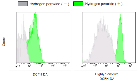
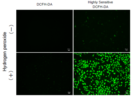
Usage examples
Detection of ROS in HeLa cells treated with hydrogen peroxide in different applications.
- HeLa cells (1×10⁴ cells/ml) in MEM (supplemented with 10% fetal bovine serum and 1% penicillin-streptomycin) were seeded into vessels and incubated for 2 nights in an incubator set at 37℃ and equilibrated with 95% air and 5% CO2.
- After the culture medium was removed, DCFH-DA or Highly Sensitive DCFH-DA Dye working solution was added.
The cells were then cultured for 30 minutes as in step 1 for 30 minutes. The cells were washed twice with HBSS. - Overall, 100 μmol/l hydrogen peroxide in HBSS was added to each well, and the cells were again cultured for 30 minutes as in step 1.
- The cells were washed twice with HBSS, and vessels were refilled with HBSS.
- Fluorescence signals were measured by a plate reader, a flow cytometer, and imagings were performed with fluorescence microscopy.
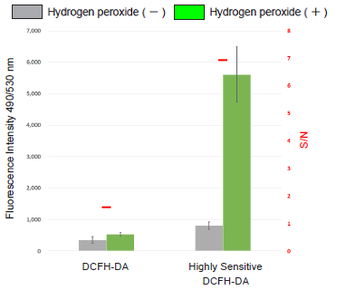
Figure 1. Fluorescence signals of HeLa cells
using a fluorescence plate reader
Detection: FITC filter
Figure 2. Fluorescence signals of HeLa cells
using a flow cytometer
Detection: GFP filter
Figure 3. Fluorescence imaging of
HeLa cells under a fluorescence microscope
Detection of endogenous ROS in RAW264.7 cells treated with lipopolysaccharide (LPS).
- RAW264.7 cells (2×10⁴ cells/ml) in DMEM (supplemented with 10% fetal bovine serum and 1% penicillin-streptomycin) were seeded on ibidi 8-well plates and cultured overnight in an incubator set at 37℃ and equilibrated with 95% air and 5% CO2.
- Medium was removed and DMEM containing LPS (500 ng/ml) was added to the cells.
- The cells were cultured for 20 hours as in step 1.
- After removing the supernatant, the cells were washed twice with HBSS.
- The cells were treated with Highly Sensitive DCFH-DA working solution and incubated for 30 minutes as in step 1.
- Supernatant was removed, and cells were washed twice with HBSS and refilled with HBSS.
- The cells were observed under a fluorescence microscope.

Figure 4. Fluorescence imaging of endogenous ROS in RAW264.7 cells
Reference
- Li J., Cell Death. Dis., 2020, 88, 11
Frequently Asked Questions / Reference


R252: ROS Assay Kit -Highly Sensitive DCFH-DA-
Revised May., 31, 2023