|
CLCC1, an ER membrane protein linked to ER ion homeostasis and ER stress control, has recently attracted attention as a factor that supports hepatic lipid homeostasis. Here, we highlight two recent papers that provide new insights into CLCC1 function. Mathiowetz et al. showed that CLCC1 directs hepatic neutral lipids toward either cytosolic lipid droplets or ER luminal lipoprotein-like particles, and reported that CLCC1 deficiency causes accumulation of giant luminal particles together with decreased nuclear pore numbers, implicating CLCC1 in ER and nuclear envelope control. Wu et al. emphasized that CLCC1 senses ER bilayer imbalance and cooperates with TMEM41B-dependent scrambling, and demonstrated that disruption of this control collapses apoB lipidation and VLDL secretion, leading, as in Mathiowetz et al., to lipid accumulation within the ER. Together, these findings provide a new mechanism for CLCC1-mediated control of neutral lipid flux. |
||||||||||||||||||||||||
|
CLCC1 promotes hepatic neutral lipid flux and nuclear pore complex assembly (Nature, 2026) Highlighted technique: In this study, Lipi-Blue or BODIPY 493/503 staining was used to visualize and quantify neutral lipid/lipid droplet phenotypes after perturbing target genes. To assess whether the giant structures formed upon CLCC1 loss were canonical cytosolic lipid droplets, Lipi-Blue staining was combined with PLIN2 as a lipid-droplet coat marker (PLIN2-positive LDs) for analysis. |
||||||||||||||||||||||||
|
CLCC1 governs ER bilayer equilibration to maintain lipid homeostasis (Nature, 2026) Highlighted technique: They quantified ER phospholipid scrambling in intact cells by adding alkyne-choline to the medium for cellular uptake and synthesis of alkyne-containing phosphatidylcholine, alkyne-PC, thereby introducing a “handle” on phospholipids. Next, they selectively permeabilized the cholesterol-rich plasma membrane with digitonin, labeled only alkyne-PC remaining on the cytosolic ER leaflet via a fluorophore–azide click reaction, and used fluorescence intensity, with higher signal indicating reduced scrambling, to assess scrambling activity. |
||||||||||||||||||||||||
Lipid Metabolism, Mitochondria and Lysosome Indicators (click to open/close)
|
||||||||||||||||||||||||
Application Note (click to open/close)
|
||||||||||||||||||||||||
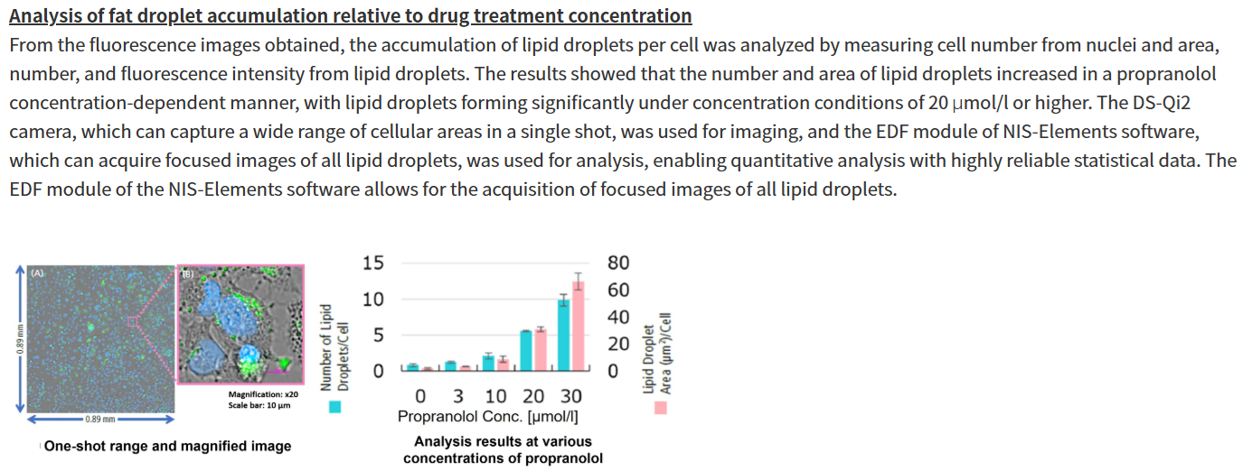
|
Propranolol (a sympathetic β-receptor blocker) was added to a human hepatocellular carcinoma cell line (HepG2 cells), and changes in lipid droplets were observed under a fluorescence microscope. The accumulation of lipid droplets was analyzed by measuring the number, area, and fluorescence intensity of lipid droplets from the acquired microscopic images.
For details of staining and analysis methods, please refer to "APPLICATION NOTE: Hepatotoxicity test of drug-induced lipidosis using high-content imaging" by Nikon Corporation. |