
Lysosomal Damage and Repair Mechanisms


Previous Science Note  

Understanding lysosomal damage and repair is key to linking cellular homeostasis with dysfunction and disease. Recent studies show that cells can respond differently depending on whether the lysosomal membrane is only stretched or is actually damaged.
The first study revealed how a damage signal is passed on to the proteins that repair the lysosomal membrane. The second showed that a lipid-transfer protein can respond even before the membrane breaks and then help deliver membrane lipids from the endoplasmic reticulum, or ER, to damaged lysosomes.
Together, these studies show that lysosomal damage should be viewed as a series of steps, including membrane stress, damage sensing, repair, functional recovery, and the cellular effects that follow when repair is insufficient.

LASER couples damage sensing to ESCRT assembly for lysosome repair
(Goul et al., Nature, 2026)

Summary
The researchers identified a protein that moves to damaged lysosomes. LC3 and related proteins, which are widely known for their roles in autophagy, are added to damaged lysosomal membranes and act as signals that attract this protein. The recruited protein then forms a complex that brings membrane-repair proteins to the damaged lysosome, linking damage sensing directly to membrane repair. In cells carrying a neurological disease-associated mutation, membrane-repair proteins were not efficiently recruited and lysosomal membrane repair was impaired. In human iPSC-derived neurons, weakening this repair pathway also increased the number of damaged lysosomes. These findings show that protecting lysosomes requires not only detecting damage, but also correctly passing that information to the membrane-repair machinery. Failure of this process may therefore contribute to neurological disease.

Highlighted technique
To identify factors involved in lysosomal repair, the researchers used large-scale gene screening and tracked repair-related proteins at damaged lysosomes by live-cell imaging and fluorescence staining. They also used fluorescent indicators of membrane leakage and resealing to determine whether damaged lysosomal membranes were successfully repaired, linking protein recruitment directly to membrane recovery.

When lysosomal repair is impaired, damaged lysosomes can remain inside the cell. Evaluating cell viability and cytotoxicity together with lysosomal damage can therefore help determine whether impaired repair has progressed to broader cellular dysfunction. 


VPS13C/PARK23 initiates lipid transfer and membrane remodeling for efficient lysosomal repair
(Adeosun et al., Nature Communications, 2026)


Summary
This study showed that a lipid-transfer protein involved in lysosomal repair responds differently to membrane stretching and actual membrane damage. The protein moved to lysosomes even before clear membrane rupture and, after damage, helped establish contact with the ER and support lipid delivery.
By tracking phosphatidylcholine, a major membrane lipid, the researchers showed that ER-derived lipids were supplied to damaged lysosomes. When this repair pathway was disrupted, lipid delivery was strongly reduced and recovery of the acidic lysosomal environment was delayed. These findings show that efficient lysosomal repair requires not only damage sensing, but also lipid supply and restoration of lysosomal function after damage.

Highlighted technique
To evaluate lysosomal repair, the researchers tracked how a lipid-transfer protein responds to membrane stress, followed phosphatidylcholine movement from the ER to damaged lysosomes, and monitored recovery of lysosomal acidity after transient damage. Together, these experiments linked damage sensing, lipid supply for membrane repair, and recovery of lysosomal function within the same repair process.

When lysosomes cannot be successfully repaired, severely damaged lysosomes can instead be removed through lysophagy, a selective form of autophagy. Therefore, by evaluating not only lysosomal pH and lysosomal mass, but also autophagic flux, we can gain a more comprehensive understanding of the cellular processes involved in lysosomal damage and repair, as well as the removal of damaged lysosomes. 

Lysosomal Related Indicators (click to open/close)
Target Kit & Probes
Lysosomal Function Analysis Kit Lysosomal Acidic pH Detection Kit - Green/Red and Green/Deep Red
High Specific Lysosommal Detection LysoPrime Green / Deep Red
Lysosomal Acidic pH Detection pHLys Red
First-time Autophagy Research Autophagic Flux Assay Kit
Autophagy detection  DAPGreen, DAPRed (Autophagosome detection), DALGreen (Autolysosome detection
Intracellular lipid radical detection Lipid Radical Probe -NBD-Pen-
Lysosomal lipid radical detection Lysosomal Lipid Radical Probe -Lyso-NBD-Pen-
Lipid Peroxidation Assay Lipid Peroxidation Probe -BDP 581/591 C11-
Application Note (click to open/close)   > Accurate Measurement for Lysosomal pH changes
 

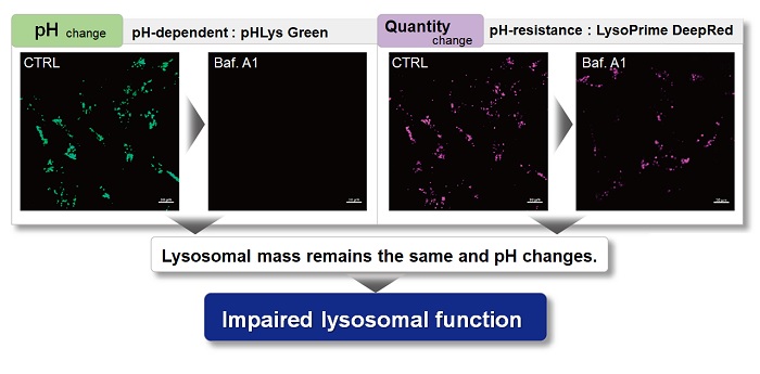
With existing reagents, it was difficult to determine whether lysosomal mass or their function (pH) fluctuated because the discussion was based on changes in the fluorescence brightness of a single dye. This kit contains pHLys Green, which is highly specific to lysosomes and shows pH-dependent changes in fluorescence, and pH-resistant LysoPrime Deep Red. Using these two dyes, lysosomal pH and volume of the same sample can be measured for a detailed analysis of lysosomal function.

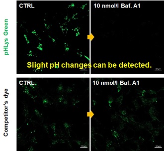
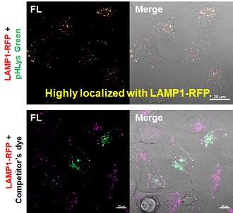
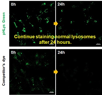
 Existing lysosomal pH detection reagents have issues with dye localization, pH sensitivity, and retention. pHLys Green is a dye that solves these issues. The improved dye retention and localization enable detection of normal lysosomes, and the improved pH sensitivity enables detection of slight pH changes.
1. High sensitive pH detection
Comparison of pH response of cells treated with low concentrations of lysosomal acidification inhibitor Bafilomycin A1
2. High specificity for lysosomes
Comparison of specificity for lysosomes using lysosomal marker protein LAMP1-GFP expressing cells
3. High retention in lysosomes
Comparison of intracellular retention

Product in Use:
   - Lysosomal Acidic pH Detection Kit-Green/Deep Red

Related Product:
   - Lysosomal Acidic pH Detection Kit-Green/Red
   - LysoPrime Deep Red - High Specificity and pH Resistance
   - pHLys Red - Lysosomal Acidic pH Detection

Product Classification

Product Classification