|
Lysosomal dysfunction is broadly linked to diseases and aging, and recent work indicates that lysosome positioning also contributes to these states. Changes in localization can influence how cells respond to stress or remodel their surrounding environment. Recent studies highlight this idea: metastatic melanoma cells shift lysosomes to the periphery to enhance invasion, while senescent cells require TIGAR-dependent perinuclear accumulation of lysosomes to maintain SASP. These findings expand the view of lysosomes beyond degradative organelles, emphasizing that their spatial organization can actively shape distinct cellular phenotypes. |
||||||||||||||||||||||
|
1. Peripheral positioning of lysosomes supports melanoma aggressiveness (Nature Communications, 2025) Summary: This study demonstrates that metastatic melanoma cells reposition lysosomes toward the cell periphery, a spatial reorganization that enhances lysosomal exocytosis, extracellular matrix degradation, and invasive capacity. By forcibly relocating lysosomes back to the perinuclear region, the authors show that invasiveness and metastatic behavior are significantly reduced, establishing lysosome positioning as a causal regulator of melanoma aggressiveness. Highlighted technique: To test whether lysosome positioning influences melanoma cell behavior, the study used an FKBP–FRB dimerization system to chemically induce binding between LAMP1-tagged lysosomes and a perinuclear motor protein. This method forces lysosomes to relocate from the cell periphery to the perinuclear region, allowing the authors to assess resulting changes in invasive and metastatic behavior. Lysosome localization was visualized by tagging LAMP1 with a fluorescent protein. Related technique Lysosomal Function Analysis, Cell Membrane Staining |
||||||||||||||||||||||
|
Summary: Senescent cells exhibit a pro-inflammatory SASP, for which proper perinuclear positioning of lysosomes is critical, but the mechanism controlling this lysosome localization had remained unclear. This study shows that TIGAR is a new key regulator that drives lysosomes to the perinuclear region, and that loss of TIGAR impairs this perinuclear repositioning and markedly reduces SASP, even though cellular senescence itself still occurs. Related technique Cellular Senescence Detection, Autophagic Flux Assay |
||||||||||||||||||||||
All Related Techniques (click to open/close)
|
||||||||||||||||||||||
Application Note (click to open/close)
|
||||||||||||||||||||||
|
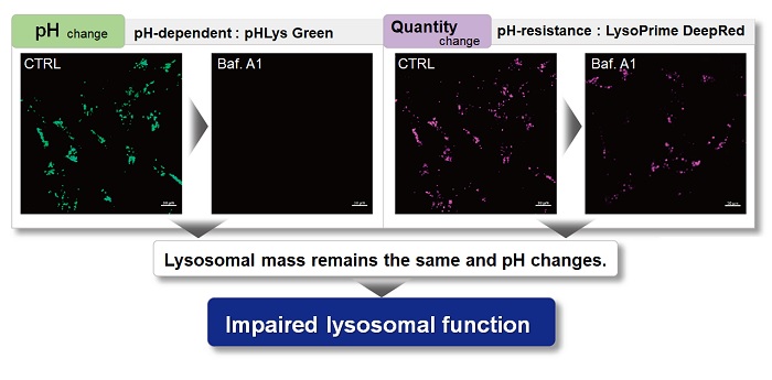
With existing reagents, it was difficult to determine whether lysosomal mass or their function (pH) fluctuated because the discussion was based on changes in the fluorescence brightness of a single dye. This kit contains pHLys Green, which is highly specific to lysosomes and shows pH-dependent changes in fluorescence, and pH-resistant LysoPrime Deep Red. Using these two dyes, lysosomal pH and volume of the same sample can be measured for a detailed analysis of lysosomal function.
|
|||
| Existing lysosomal pH detection reagents have issues with dye localization, pH sensitivity, and retention. pHLys Green is a dye that solves these issues. The improved dye retention and localization enable detection of normal lysosomes, and the improved pH sensitivity enables detection of slight pH changes. | |||
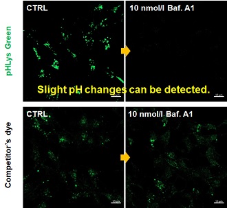
| 1. High sensitive pH detection Comparison of pH response of cells treated with low concentrations of lysosomal acidification inhibitor Bafilomycin A1 |
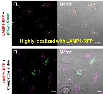
2. High specificity for lysosomes Comparison of specificity for lysosomes using lysosomal marker protein LAMP1-GFP expressing cells |
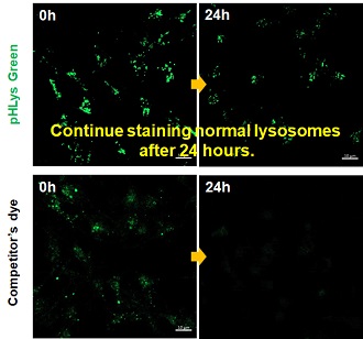
3. High retention in lysosomes Comparison of intracellular retention |
|
 |
 |
 |
|
|
Product in Use: Related Product: |
|||