Ferroptosis Control Through Lysosomal Iron Homeostasis [May 20, 2025] 

Previous Science Note

Ferroptosis is influenced by lysosomal activity, particularly through its roles in iron homeostasis, membrane integrity, and the breakdown of antioxidant defenses. This Science Note introduces recent insights into how lysosomal function influences ferroptotic cell death, highlighting emerging mechanisms linking lysosomal metabolism, iron release, and lipid peroxidation.

Activation of lysosomal iron triggers ferroptosis in cancer (Nature, 2025)
Summary: This study shows that the ferroptosis inhibitor Liproxstatin-1 localizes to lysosomes and suppresses iron-dependent lipid peroxidation, identifying lysosomal iron as a key trigger of ferroptosis. Additionally, the authors developed Fento-1, which selectively activates lysosomal iron, and demonstrated that controlling lysosomal iron could serve as a therapeutic strategy.

Highlighted technique: Click chemistry, exemplified by azide–alkyne cycloaddition, enables rapid and selective covalent linkage between functional groups. This study used an alkyne-containing Liproxstatin-1 analogue (cLip-1) and visualized its intracellular distribution via click reaction to clarify Liproxstatin-1’s mechanism of action.

 Related technique  Intracellular Iron Detection, Lipid Peroxide Detection (used in this article)

SLC7A11 is an unconventional H+ transporter in lysosomes (Cell, 2025)
Summary: This study identifies lysosomal xCT (SLC7A11) as a previously unrecognized mediator of slow proton leak via cystine/glutamate exchange, revealing a novel mechanism for regulating lysosomal pH. Inhibition of SLC7A11 leads to lysosomal hyper acidification, impaired degradation, ferroptosis, and α-synuclein aggregation.

Highlighted technique: The authors screened a lysosomal membrane protein KO library by measuring lysosomal acidity with a pH-sensitive dye. Unlike most cells, SLC7A11-KO cells maintained acidity after bafilomycin A1 treatment, identifying SLC7A11 as a key regulator of lysosomal H⁺ efflux.

 Related technique  Lysosomal Analysis, Cystine Uptake Assay

Glucose starvation causes ferroptosis-mediated lysosomal dysfunction (iScience, 2024)
Summary: Glucose starvation decreases lysosomal protein expression and causes lysosomal damage, leading to ferroptosis through iron accumulation. While GPX4 accumulates on lysosomes to suppress ferroptosis, the inactivation of other lysosome-associated enzymes contributes to dysfunction and cell death.

Highlighted technique: In this study, lysosomal functional changes under glucose starvation were evaluated using pH-sensitive probes, protein analysis from isolated lysosomal fractions and autophagy assay. Imaging of lipid peroxidation and intracellular iron was further used to link glucose deprivation with lysosomal dysfunction and ferroptosis.

 Related technique  Intracellular Iron Detection (used in this article),  Autophagy Detection (used in this article)

Related Techniques (click to open/close)
Target Kit & Probes
Ferroptosis Indicator: ferrous ion (Fe2+) FerroOrange(intracellular), Mito-FerroGreen(mitochondria)
Ferroptosis Indicator: lipid peroxidation Liperfluo(intracellular), MitoPeDPP(mitochondria)
Lipid Peroxidation Assay Lipid Peroxidation Probe -BDP 581/591 C11-
Lysosomal function Lysosomal Acidic pH Detection Kit -Green/Red and Green/Deep Red
First-time autophagy research Autophagic Flux Assay Kit
Cystine Uptake detection Cystine Uptake Assay Kit
Glutamate detection Glutamate Assay Kit-WST
Glutathione Quantification GSSG/GSH Quantification Kit
Total ROS detection Highly sensitive DCFH-DA or Photo-oxidation Resistant DCFH-DA
Cell proliferation/ cytotoxicity assay Cell Counting Kit-8 and Cytotoxicity LDH Assay Kit-WST
Application Note (click to open/close)
  > When Lysosomes Go Neutral: Iron Loss Unveiled

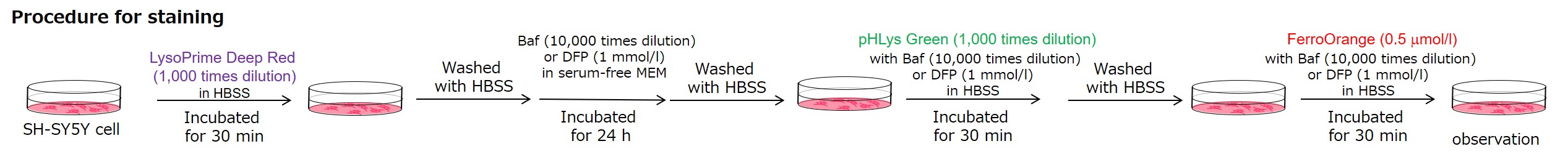
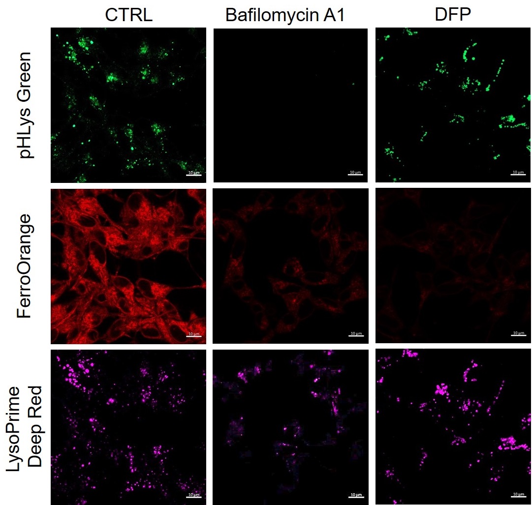
In neurodegenerative diseases, the relationship between lysosomal function and iron has attracted attention, and it has been reported* that lysosomal neutralization prevents the breakdown of iron stores (Transferrin or Ferritin), resulting in a decrease in intracellular iron.   *Mol Cell., 202077(3), 645-655


Lysosomal pH changes and intracellular iron changes in the same sample were detected using SH-SY5Y cells supplemented with lysosomal acidification inhibitor (Bafilomycin A1) or iron chelator (Deferipron (DFP)). (Lysosomal pH: Lysosomal Acidic pH Detection kit - Green/Deep Red, Intracellular iron: FerroOrange [Code:F374])
The results showed that the addition of Bafilomycin A1 decreased the fluorescence of FerroOrange, confirming the decrease in intracellular iron. The fluorescence of LysoPrime DeepRed remained almost unchanged, while the fluorescence of pHLys Green decreased due to lysosomal neutralization. These results suggest that there is a relationship between changes in intracellular iron and lysosome function.

<Condition>
pHLys Green (Green) : Ex=488 nm, Em=486-574 nm
FerroOrange (Red) : Ex=561 nm, Em=550-650 nm
LysoPrime Deep Red (Violet) : Ex=633 nm, Em=599-700 nm


Product Classification

Product Classification