Ferroptotic Cells Trigger Nearby Cell Death [Apr. 15, 2025]

Previous Science Note

Ferroptotic cells release signals that spread beyond themselves, inducing lipid peroxidation and death in neighbouring cells. This Science Note introduces recent advances in our understanding of how signals from ferroptotic cells spread and the molecular mechanisms underlying this process.

Ferroptosis spreads to neighboring cells via plasma membrane contacts 
(Nature Communications, 2025)

Ferroptosis spreads to neighbouring cells through the transfer of lipid peroxidation across closely adjacent plasma membranes. This spread of lipid peroxidation is dependent on the presence of extracellular iron.

Highlighted technique: In this study, the authors developed a novel model for ferroptosis induction by creating cells in which the lipid peroxide-detoxifying enzyme GPX4 can be selectively depleted by optogenetic activation. This system allowed the induction of ferroptosis in specific cells, allowing the observation of lipid peroxidation and the spread of ferroptosis to neighbouring cells.

 Related technique  Lipid Peroxidation Detection, Intracellular Iron Detection

Emergence of large-scale cell death through ferroptotic trigger waves
(Nature, 2024)

This study shows that ferroptosis propagates as a trigger wave at a constant speed (~5.5 μm/min) over distances ≥5 mm, driving large-scale cell death seen in development and disease. ROS released during ferroptosis spread to neighboring cells and induce further death, sustained by positive feedback loops that amplify ROS production.

Highlighted technique: The propagation of ferroptosis was analysed by dynamic observation using a combination of intracellular ROS staining and dead cell staining. Real-time time-lapse fluorescence microscopy revealed that ROS generation from ferroptotic cells preceded the wave-like spread of cell death.

 Related technique  Highly Sensitive ROS Detection Dye, Long-Term ROS Detection Dye

Microglial ferroptotic stress causes non-cell autonomous neuronal death
(Molecular Neurodegeneration, 2024)

Ferroptotic stress in microglia initiates an inflammatory cascade that promotes the transformation of astrocytes into a neurotoxic state, ultimately leading to neuronal death. This pathway highlights a mechanism by which localised oxidative stress in glial cells can contribute to wider neurodegeneration.

Highlighted technique: In this study, ferroptosis inducers were applied to microglia, astrocytes or mixed glial cultures and the resulting conditioned media were added to neurons to assess lipid peroxidation and cell viability. Neuronal death was only observed with media from ferroptosis-treated mixed glial cultures, indicating that neurotoxicity requires interaction between multiple glial cell types.

 Related technique  Lipid Peroxidation Assay, Cell Viability Assay

Related Techniques (click to open/close)
Target Kit & Probes
Ferroptosis Indicator: ferrous ion (Fe2+) FerroOrange(intracellular), Mito-FerroGreen(mitochondria)
Ferroptosis Indicator: lipid peroxidation Liperfluo(intracellular), MitoPeDPP(mitochondria)
Lipid Peroxidation Assay Lipid Peroxidation Probe -BDP 581/591 C11-
Total ROS detection Highly sensitive DCFH-DA or Photo-oxidation Resistant DCFH-DA
Glutathione Quantification GSSG/GSH Quantification Kit
Cystine Uptake detection Cystine Uptake Assay Kit
MDA detection MDA Assay Kit
Mitochondrial superoxide detection MitoBright ROS Deep Red - Mitochondrial Superoxide Detection
Apoptosis detection in multiple samples Annexin V Apoptosis Plate Assay Kit
Cell proliferation/ cytotoxicity assay Cell Counting Kit-8 and Cytotoxicity LDH Assay Kit-WST
Application Note (click to open/close)
  > Erastin-Induced Ferroptosis: Evaluating Intracellular Uptake and Redox Balance

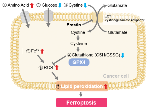
We investigated the transition of cellular metabolisms in A549 cells treated with erastin, a known ferroptosis inducer. Our results revealed the following.

Results
- The inhibition of cystine uptake by erastin led to a depletion of cysteine, which in turn increased the compensatory uptake of other amino acids.
- Glucose uptake, which typically promotes ferroptosis*, was found to decrease upon erastin treatment, suggesting a potential cellular self-defense mechanism.
- The depletion of cysteine resulted in a decrease in glutathione levels and an increase in Fe2+, ROS, and lipid peroxides, all of which are recognized markers of ferroptosis.

  Cell Line: A549
  Incubation Conditions: 100 μmol/l Erastin/MEM, 37℃, 3h
  *Reference: Xinxin Song, et al., Cell Reports, (2021)

Products in Use
① Amino Acid Uptake: Amino Acid Uptake Assay Kit
② Glucose Uptake: Glucose Uptake Assay Kit-Green
③ Cystine Uptake: Cystine Uptake Assay Kit
④ Intracellular glutathione: GSSG/GSH Quantification Kit
⑤ Intracellular labile Fe: FerroOrange
⑥ Intracellular total ROS: ROS Assay Kit -Highly Sensitive DCFH-DA-
⑦ Lipid Peroxides: Liperfluo


Product Classification

Product Classification