|
Autophagosome formation alone does not show whether cargo reaches lysosomes, is degraded, or remains linked to stress. In a neurodegeneration model, defects in EPG5, the autophagy-tethering factor, were associated with impaired autophagosome-lysosome fusion, defective mitochondrial clearance, Ca2+ overload, cytosolic mtDNA, and innate immune activation. In glioblastoma, cells with high lysosomal activity showed increased mitochondrial membrane potential, mitochondrial ROS, oxygen consumption rate, and tolerance to temozolomide, a DNA-damaging drug for glioblastoma treatment. Together, these findings support assessing fusion with lysosomes, cargo degradation, and stress readouts when evaluating autophagy in disease models.
|
 |
Pathogenic variants in the autophagy-tethering factor EPG5 drive neurodegeneration through mitochondrial dysfunction and innate immune activation
(Singh et al., Nature Communications, 2026)
Summary:
This study investigated how EPG5-related impairment of autophagosome-lysosome fusion links defective mitochondrial clearance to calcium stress and inflammatory signaling in neurodegeneration. In patient-derived fibroblasts and iPSC-derived cortical neurons, defective autophagy was associated with reduced mitochondrial membrane potential, calcium overload, mitochondrial DNA release into the cytosol, and activation of innate immune signaling.
Highlighted technique:
To assess mitochondrial dysfunction downstream of EPG5-related autophagosome-lysosome fusion impairment, the authors measured membrane potential, oxygen consumption, mitochondrial respiratory protein levels, mitochondrial ROS, and cytosolic and mitochondrial calcium responses in patient-derived fibroblasts and iPSC-derived cortical neurons.
Mitochondrial dysfunction can be evaluated by membrane potential, ROS. A mitochondrial fractionation kit enables collection of intact mitochondria from tissue samples for direct assessment of mitochondrial function, including OCR and Complex I activity.
Lysine-arginine imbalance overcomes therapeutic tolerance governed by the transcription factor E3-lysosome axis in glioblastoma
(Jing et al., Nature Communications, 2025)
Summary:
This study examined whether lysosomal function could indicate malignant states in glioblastoma (GBM) that are not fully explained by genetic background alone. Cells with higher lysosomal activity showed greater sphere-forming capacity, mitochondrial membrane potential, mitochondrial ROS production, and oxygen consumption rate. The lysosome-regulating transcription factor TFE3 linked lysosomal function to mitochondrial activation and tolerance to temozolomide, a standard GBM therapy.
Highlighted technique:
To examine whether lysosomal activity was coupled to mitochondrial function in patient-derived GBM cells, the authors inhibited lysosomal function with bafilomycin A1 and measured mitochondrial membrane potential, mitochondrial ROS, and oxygen consumption rate to assess mitochondrial energetics and respiratory capacity.
Fluorescent reagents enable live-cell detection of autophagosomes, autolysosomes, mitophagy, and lysosomes without genetic modification. Lysosome probes are available as pH-resistant or pH-dependent options.
 
Autophagy and Mitochondria Related Activity Indicators (click to open/close)
| Target |
Kit & Probes |
| First-time Autophagy Research |
Autophagic Flux Assay Kit |
| Lysosomal Function Analysis Kit |
Lysosomal Acidic pH Detection Kit -Green/Red and Green/Deep Red |
| High Specific Lysosomal Detection |
LysoPrime Green / Deep Red |
| Lysosomal Acidic pH Detection |
pHLys Red |
| Mitophagy detection |
Mitophagy Detection Kit |
| Intact Mitochondria Fractionation |
IntactMito Fractionation Kit for Tissue |
| Oxygen consumption rate assay |
Extracellular OCR Plate Assay Kit |
| MitoComplex-I Activity Assay |
MitoComplex-I Activity Assay Kit |
| Mitochondrial Staining |
MitoBright LT Green / Red / Deep Red |
| Mitochondrial membrane potential detection |
JC-1 MitoMP Detection Kit, MT-1 MitoMP Detection Kit |
| Mitochondrial superoxide detection |
MitoBright ROS Deep Red - Mitochondrial Superoxide Detection |
|
Application Note I (click to open/close)
> Accurate Autophagy Imaging with Lysosome Staining
|
|

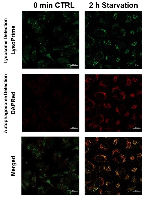
We performed fluorescence imaging under amino acid starvation in HeLa cells stained with the autophagosome detection probe DAPRed (Code: D677) and LysoPrime Green (Code: L261). The fluorescence signal of LysoPrime Green increased, and lysosomal localization was confirmed over time. These results indicate that the co-localization rate with DAPRed fluorescence is sufficient, enabling accurate autophagy analysis.
|
|
|
Application Note II (click to open/close)
> Induction of Mitophagy in Parkin Expressed HeLa cells
|
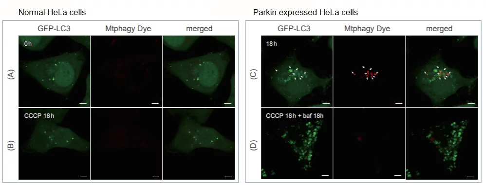
CCCP(carbonyl cyanide m-chlorophenyl hydrazone) has been added to normal and Parkin expressed cells. The strong fluorescence was not observed in normal HeLa cells(A)(B). On the other hand, the trong fluorescence was shown in Parkin expressed cells in 18 hours after additon of CCCP(C). Some of the puncta are co-localized with the autophagy marker(GFP-LC3). In addition, suppressed fluorescence of Mtphagy dye was observed when autophagy inhibitor, bafilomycin was added to Parking expressed cells(D). Because lysosomal pH was increased by the additon of bafilomycin.
|
|
|