Previous Science Note
| Autophagy is increasingly recognized as a key regulator in disease pathogenesis, influencing the mechanisms of protein aggregation, secretion, and cellular clearance. This Science Note introduces three studies highlighting how autophagy suppression by PHGDH, the secretion mechanism of the antioxidant protein PARK7, and p97/VCP-dependent aggrephagy contribute to the progression of neurodegeneration and cancer. | ||||||||||||||||||||||
|
Transcriptional regulation by PHGDH drives amyloid pathology in Alzheimer’s disease (Cell, 2025) Highlighted technique: This study demonstrated that PHGDH increases the expression of IKKα and HMGB1, which are involved in the suppression of autophagy via the mTOR pathway, at both the mRNA and protein levels in brain organoid astrocytes. Impaired autophagy was also confirmed by a reduction in autophagosomes using fluorescent small-molecule probes. Related technique Autophagic Flux Detection, Apoptosis Detection |
||||||||||||||||||||||
|
Unconventional secretion of PARK7 requires lysosomal delivery via chaperone-mediated autophagy and specialized SNARE complex (PNAS, 2025) Highlighted technique: In this study, autophagy was monitored by expressing LC3 fused with either GFP or mRFP in cells. While both GFP and mRFP fluorescence are visible in autophagosomes, only mRFP remains detectable in autolysosomes due to the quenching of GFP in the acidic environment. Related technique Autophagic Flux Detection Lysosome function |
||||||||||||||||||||||
|
p97/VCP is required for piecemeal autophagy of aggresomes (Nature Communications, 2025) Highlighted technique: In this study, retinoic acid-differentiated SH-SY5Y cells, which mimic neuron-like properties and are commonly used to study aggresomes, were utilized as a disease-relevant model. Autophagy was assessed by immunofluorescence staining of LC3, revealing that p97/VCP inhibition blocked aggresome clearance. Related technique Autophagic Flux Detection Mitophagy Detection |
||||||||||||||||||||||
Related Techniques (click to open/close)
|
||||||||||||||||||||||
Application Note I (click to open/close)
|
||||||||||||||||||||||
|
|
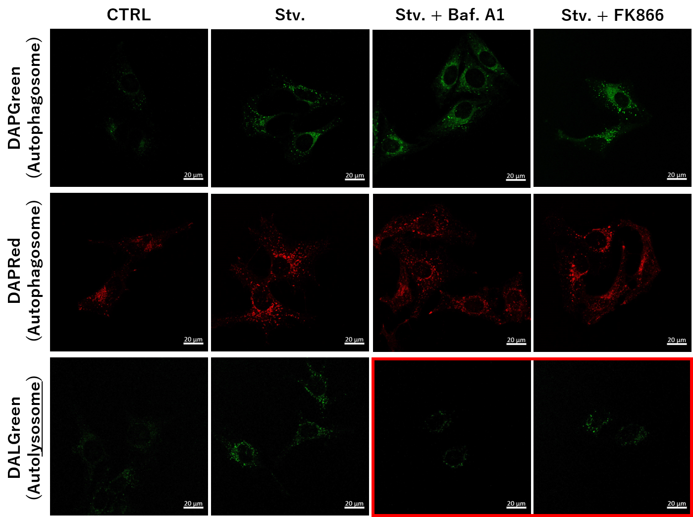
We determined how FK866-induced lysosomal deacidification affects the autophagy-lysosomal pathway. After staining with DAPGreen/DAPRed (for detecting autophagosome), or DALGreen (for detecting autolysosome), HeLa cells were starved in HBSS incubation and then treated with FK866 or Bafilomycin A1. Under the starvation condition, the fluorescent signals from all dyes increased, indicating the proceeding autophagy-lysosomal pathway. On the other hand, only DALGreen's signals were decreased in FK866 and Bafilomycin A1 treated cells with starvation conditions. These results clearly suggested that FK866 inhibits the autophagy-lysosomal pathway by NAD+ depletion-induced lysosomal deacidification. Nampt inhibitor, FK866 inhibits the progress of autophagosome to autolysosome by lysosomal deacidification. A recent finding shows that the dysfunctional condition of nicotinamide adenine dinucleotide (NAD+) biosynthetic enzyme, Nampt induces lysosomal deacidification1). In this section, we tried to determine how NAD+ depletion-induced lysosomal deacidification affects the autophagy-lysosomal pathway. 1) Mikako Yagi, et. al., EMBO J., 40(8), e105268 (2021) |
Application Note II (click to open/close)
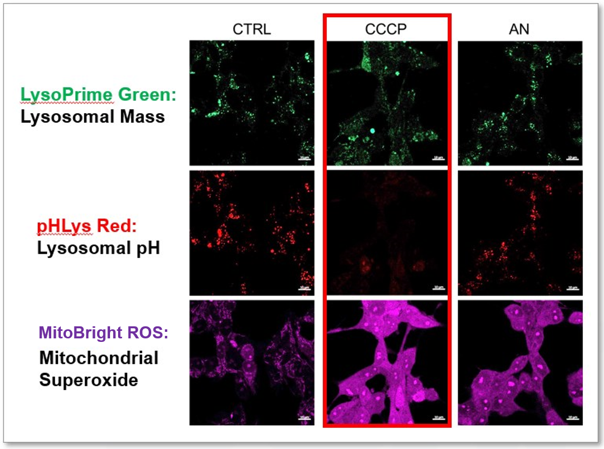
> Simultaneous Detection of Lysosomal and Mitochondrial Dysfunction
|
|
We tried the simultaneous detection of lysosomal and mitochondrial dysfunction in Hela cells treated with CCCP or Antimycin (AN). CCCP and AN are well-known inducers of mitochondrial ROS regarding loss of mitochondrial membrane potential. Recent research showed the result that CCCP induces not only mitochondrial ROS but also lysosomal neutralization. To detect mitochondrial ROS, HeLa cells were labeled by MitoBright ROS Deep Red - Mitochondrial Superoxide Detection, and the lysosomal mass and pH were detected separately with LysoPrime Green and pHLys Red. Co-staining with MitoBright ROS and Lysosomal dyes demonstrated that CCCP causes lysosomal neutralization and mitochondrial ROS induction at the same time. Products in Use
|