Autophagy for Cellular Homeostasis

 [Aug. 20, 2024] Previous Science Note

Cell death and the dysfunction of autophagy and lysosomes are closely linked in maintaining cellular health. When autophagy is impaired, cells cannot effectively degrade and recycle damaged components, leading to the accumulation of toxic materials and cellular stress. Lysosomal dysfunction exacerbates this problem, as these organelles are essential for the degradation of autophagosomal contents. The resulting cellular damage and stress can trigger cell death pathways that contribute to various diseases, including neurodegenerative disorders and cancer.

Tumor suppressor Par-4 activates autophagy-dependent ferroptosis
Click here for the original article: Karthikeyan Subburayan, et. al., Communications Biology, 2024.

Point of Interest
- Par-4/PAWR activation is critical during ferroptosis, with its depletion blocking and overexpression enhancing this ferroptosis process.

- Upregulation of Par-4 promotes NCOA4-dependent ferritinophagy, a selective autophagy process that degrades ferritin, increasing free iron release, lipid peroxidation, and ROS production.

- Par-4 inhibition blocks ferroptosis-driven tumor suppression, highlighting its potential for cancer therapy.

The aging mouse CNS is protected by an autophagy-dependent microglia population promoted by IL-34
Click here for the original article: Rasmus Berglund, et. al., Nature Communications, 2024.

Point of Interest
- A microglial population emerges in the cortical regions of aging mice, characterized by a transcriptome indicative of activated autophagy.

- This population is found to be dependent on IL-34, a ligand for CSF1R.

- Loss of autophagy-dependent microglia leads to neuronal and glial cell death and increased mortality in aged mice exposed to neuroinflammation.

- Conversely, IL-34-mediated microglial expansion is protective.

Autophagy counters inflammation-driven glycolytic impairment in aging hematopoietic stem cells
Click here for the original article: Paul Dellorusso, et. al., Cell Stem Cell, 2024.

Point of Interest
- Autophagy in old hematopoietic stem cells (oHSCs) is a protective response to chronic inflammation, preserving regenerative capacity despite impaired glycolysis.

- Inflammation suppresses glucose uptake and suppresses glycolysis in oHSCs via Socs3 inhibition of AKT/FoxO signaling, but Socs3-mediated autophagy allows metabolic compensation and a shift toward adaptive lipid metabolism.

- Fasting/refeeding restores the glycolytic state and enhances the regenerative potential of oHSCs.

Related Technique in This Topic 

First-time autophagy research
Autophagic Flux Assay Kit
Autophagy detection
DAPRed (Autophagosome detection), DALGreen (Autolysosome detection)
Glutathione Quantification
GSSG/GSH Quantification Kit
Lysosomal function
Lysosomal Acidic pH Detection Kit-Green/Red and Green/Deep Red
Total ROS detection
Highly sensitive DCFH-DA or Photo-oxidation Resistant DCFH-DA
Cellular senescence detection
SPiDER-βGal for live-cell imaging or flow cytometry / microplate reader / tissue samples.
Glycolysis/Oxidative phosphorylation Assay
Extracellular OCR Plate Assay KitGlycolysis/OXPHOS Assay Kit
Glucose Uptake Capacity Assay
Glucose Uptake Assay Kit-BlueGreenRed

Related Applications

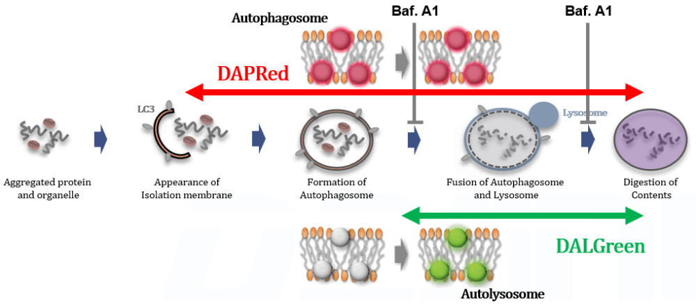
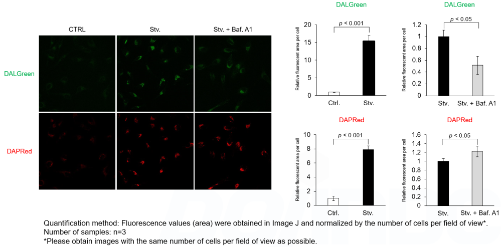
Analysis of autophagic flux without transfection

DALGreen and DAPRed labeled HeLa cells were used to evaluate changes in autophagic flux induced by the lysosomal acidification inhibitor bafilomycin A1 (Baf. A1). Compared to starvation conditions, the fluorescence signals of DALGreen were decreased under inhibited conditions of autolysosome formation by the addition of Baf. A1. In contrast, the fluorescence signals of DAPRed were increased under the same conditions, indicating that Baf. A1 led to the accumulation of autophagosome.

Experimental Data

Experimental Conditions
CTRL: Normal condition, Stv.: Induction of autophagy, Stv. + Baf. A1: Inhibition of autolysosome formation
DALGreen filter set: 488 nm (Ex), 490–550 nm (Em)
DAPRed filter set: 561 nm (Ex), 565–700 nm (Em)

Procedure

1. HeLa cells were seeded (1.0 x 10⁴ cells/well) on a μ-slide 8 well plate (ibidi) and cultured overnight at 37°C in an incubator equilibrated with 95% air and 5% CO2.

2. After washing twice with MEM containing 10% fetal bovine serum, 200 μl of DALGreen/DAPRed working solution (DALGreen: 1 µmol/l, DAPRed: 0.2 µmol/l) and the cells were incubated at 37°C for 30 minutes.

3. The supernatant was discarded, and the cells were washed twice with MEM containing 10% fetal bovine serum.

4. Samples were prepared under the following conditions.
  • MEM containing 10% fetal bovine serum (200 µl) was added to the well, and the cells were incubated at 37°C for 2 hours 20 minutes. (Control)
  • Amino acid-free medium (FUJIFILM Wako Pure Chemical Industries, Ltd., Catalogue code: 048-33575) (200 μl) was added to the well, and the cells were incubated at 37°C for 2 hours 20 minutes. (Starvation)
  • Amino acid-free medium (200 μl) was added to the well, and the cells were incubated at 37°C for 2 hours. The supernatant was discarded, bafilomycin A1 working solution (10,000 times dilution, 200 μl), an inhibitor of lysosomal acidification, was added to the well, and the cells were incubated at 37°C for 20 minutes. (Inhibition of autolysosome formation)

5. The stained cells were observed under a confocal fluorescence microscope.

Products in Use
Autophagic Flux Assay Kit


Product Classification

Product Classification