Ferroptosis Activation, Inhibition, and Sensitization

Latest Science Note
Dojindo Science Note [Feb. 13, 2024]

Ferroptosis is a form of programmed cell death characterized by the accumulation of lipid peroxides to lethal levels and is distinct from other forms of cell death such as apoptosis or necrosis. In cancer, ferroptosis can act as a double-edged sword: on the one hand, inducing ferroptosis in cancer cells may be a promising therapeutic strategy, as many cancers are resistant to traditional forms of cell death such as apoptosis. On the other hand, cancer cells can develop resistance to ferroptosis, which contributes to therapy resistance and tumor progression. Understanding and manipulating the pathways that regulate ferroptosis in cancer cells holds the potential for developing novel cancer treatments and overcoming resistance to existing therapies.

Targeted activation of ferroptosis in colorectal cancer via LGR4 targeting overcomes acquired drug resistance
Click here for the original article: Hao Zheng, et. al., Nature Cancer, 2024.

7-Dehydrocholesterol is an endogenous suppressor of ferroptosis
Click here for the original article: Florencio Porto Freitas et. al., Nature, 2024.

Sensitization of cancer cells to ferroptosis coincident with cell cycle arrest
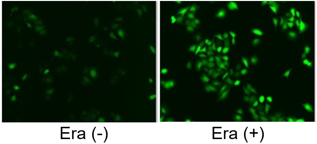
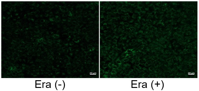
Click here for the original article: Jason Rodencal et. al., Cell Chemical Biology, 2023.

Point of Interest
- Chemoresistant cancer-derived organoids exhibited increased expression of LGR4 and activation of the Wnt signaling pathway.

- Inhibition of LGR4-Wnt signaling sensitized drug-induced ferroptosis.

- LGR4-dependent Wnt signaling upregulated SLC7A11, a key inhibitor of ferroptosis, leading to drug resistance.

Point of Interest
- High concentrations of 7-DHC are cytotoxic to developing neurons by promoting lipid peroxidation.

- On the other hand, 7-DHC accumulation confers a prosurvival function in cancer cells.

- 7-DHC effectively protects (phospho)lipids from autoxidation and subsequent fragmentation due to its superior reactivity with peroxyl radicals.

- Accumulation of 7-DHC induces a shift to a ferroptosis-resistant state in tumors.

Point of Interest
- Stabilization of p53 and inhibition of CDK4/6 enhance ferroptosis induced by GPX4 inhibitors.

- Stabilization of p53 and inhibition of CDK4/6 decrease the expression of MBOAT1 and EMP2.

- Loss of EMP2 increases cell sensitivity to GPX4 inhibitors by altering lipid metabolism.

- An orally bioavailable GPX4 inhibitor shows in vivo activity.

Related Techniques
           Intracellular lipid peroxidation measurement Liperfluo
           Mitochondria lipid peroxidation measurement MitoPeDPP
           Intracellular ferrous ion (Fe2+) detection FerroOrange
           Mitochondria ferrous ion (Fe2+) detection Mito-FerroGreen
           Mitochondrial superoxide detection MitoBright ROS Deep Red - Mitochondrial Superoxide Detection
           Total ROS detection Highly sensitive DCFH-DA or Photo-oxidation Resistant DCFH-DA 
           Lipid droplets detection Lipi-Blue / Green / Red Deep Red
           Antibody/Protein labeling with fast and
           high recovery
Fluorescein, Biotin, and Peroxidase Labeling Kit - NH2
Related Applications

The simultaneous detection of lysosomal function with Mitochondrial ROS and intracellular Fe2+

Lysosomal Function and Iron Homeostasis


Recent reports suggest that lysosomal neutralization can result in iron depletion, consequently leading to the disruption of cell viability. To verify this, HeLa cells were labeled with FerroOrange for Fe2+ detection, and the lysosomal mass and pH were separately detected with LysoPrime DeepRed and pHLys Green (a product currently under development). Co-staining with FerroOrange and Lysosomal dyes demonstrated that Bafilomycin A1 (Baf. A1), an inhibitor of lysosomal acidification, causes iron depletion consistent with the findings reported in the article. Interestingly, the iron chelator, Deferiprone (DFP), did not impact lysosomal pH, suggesting that lysosomal function plays a key role in managing iron homeostasis.

Reference: Ross A Weber, et. al., Mol Cell (2020)

Products in Use
   - FerroOrange
   - pHLys Green*
   - LysoPrime Deep Red

*pHLys Green is available as the "Lysosomal Acidic pH Detection Kit-Green/Deep Red". 


Induction of Ferroptosis by Erastin

Erastin is a known inducer of ferroptosis. By inhibiting the cystine transporter (xCT), erastin inhibits the uptake of cystine. Cystine is the raw material for GSH. Therefore, Erastin ultimately decreases the amount of GSH. Decreased GSH then results in lipid peroxide accumulation and induction of ferroptosis.
The following experimental examples show changes in each aforementioned index as a consequence of erastin stimulation. Measurements are made using Dojindo reagents.

Using erastin-treated A549 cells, we measured intracellular Fe2+, ROS, lipid peroxide, glutathione, glutamate release into the extracellular space, and cystine uptake. As a result, inhibition of xCT by elastin was observed and also the release of glutamate and uptake of cystine were decreased. Furthermore, elastin treatment decreased intracellular glutathione while it increased intracellular Fe2+ , ROS, and lipid peroxides.

①Cystine Uptake        

Cystine Uptake Assay Kit

②Released Glu            

Glutamate Assay Kit-WST

③Intracellular GSH     

GSSG/GSH Quantification Kit

④Intracellular Fe2+     

FerroOrange

⑤Intracellular ROS     

Highly Sensitive DCFH-DA

⑥Intracellular Lipid   

Liperfluo

Related Selection Guide & Science Note

Ferroptosis Guide     ​Cellular Senecence     Mitochondria

Metabolism     Autophagy     Lysosome


Product Classification

Product Classification